

Hypothyroid- Source or Scapegoat? The mistaken identity of a metabolic culprit.
Dr. Richard Maurer, ND
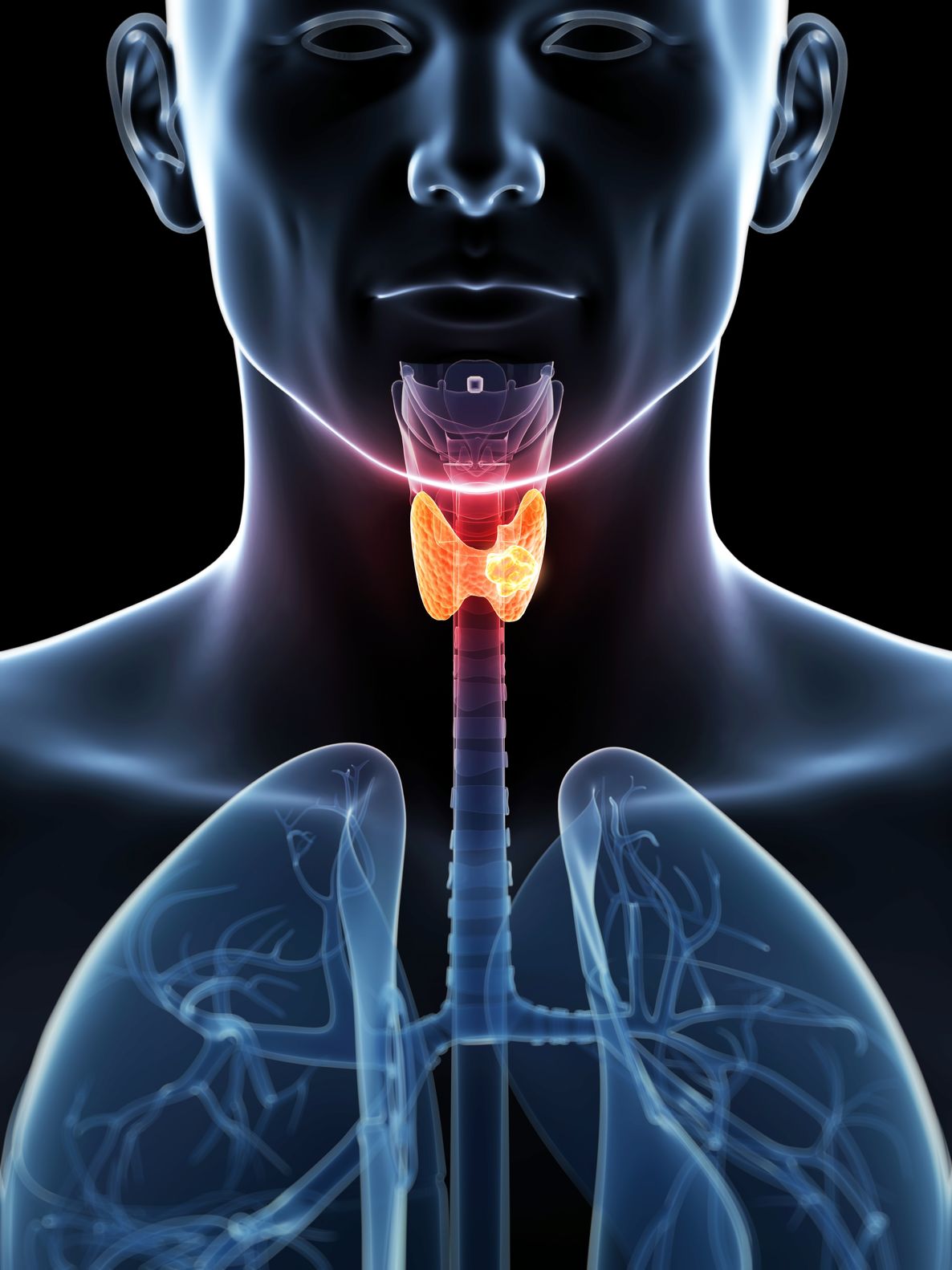
The thyroid gland has become an unruly scapegoat in the pasture of self-help and wellness. As I write this, a search on Amazon reveals 7446 books, mostly self-help, about thyroid. The book descriptions offer an alluring answer to the questions and uncertainty patients feel about their symptoms. The book descriptions state, “A thyroid problem could be the cause of your fatigue, irritability, weight problem, low libido, depression, brain fog, hair loss, poor complexion, digestive problem and so on. And since the book’s entry point is a questionnaire, it follows that the reader with any of these symptoms wants to fix the broken gland that is wreaking havoc behind the curtain of their otherwise healthy life. To blame a single set of hormones for so many symptoms is enticing, but likely false. It is up to us clinicians to rationally provide an effective solution that is true for the patient. The past decade of research has eroded the tidy line that linked the thyroid as the cause of so many effects. If the thyroid is no longer the prime cause of so much suffering, what is going on? Why is the thyroid gland erroneously blamed when she is usually doing her job, perfectly? Let’s review her job description for a moment.
Metabolism is the sum of the physical and chemical processes in an organism by which its material substance is produced, maintained, and destroyed, and by which energy is made available. Anabolism requires energy to build complex tissue and Catabolism releases energy as complex tissue is broken down into simpler components.
Humans, and most other organisms, have evolved to favor anabolism, especially when times get tough. Until recently, mortal disease was described as “consumption”. Whether during The Plague or due to tuberculosis, the victim would literally be consumed by the illness; death was a process of wasting away from starvation. Our endocrine system is brilliantly prepared to avert this pending end.
A simple downward adjustment of the idle speed of the body helps preserve precious calories, just as I use less oil to heat my house when I keep the temperature set to 59F rather than 62F. In a similar New England survival adaptation, a little insulin resistance and high insulin can help stockpile some of the calories consumed at meals. I mention this thrifty storage trend not only as a convenient comparison, but because research has revealed that these two metabolic trends are related. “Save more” and “spend less” are two sides of the same coin and it might just be that the insulin resistance trend comes first.
Human cellular metabolism is regulated by the following hormones and likely many others: Triiodothyronine – T3/ Estrogen/ Progesterone/ DHEA/ Growth Hormone/ GHIH/ Testosterone/ Cortisol/ Glucagon/ Insulin/ Leptin/ Epinephrine/ Norepinephrine/ Non-hormonal cellular cytokines play a crucial role too.
The list helps display that Triiodothyronine (T3), is just one of many metabolic regulators. Active metabolism, generated by exertion, stress, fear, and illness, engages every hormone and cytokines in the above tally. And of course these compounds all work at rest too, but T3 has been generously given the lion’s share of credit for the body’s basal (resting) metabolic rate (BMR). Let’s go back to the book that claims that a thyroid problem is causing the resistant weight gain, fatigue, insomnia, etc. We should run a test for this right?
The thyroid gland produces mostly (80%) thyroxine (T4) and the remainder as Triiodothyronine (T3). T4 is a prohormone with only about 20% of the metabolic activity in the cells compared to T3. Importantly, 98% of the T3 measured in the blood is due to spillover from the activation step in the peripheral tissue, not from thyroid gland production.
For decades the metric for adequate thyroid function has followed a logical, albeit imperfect, flow from one hormone to another: The diagram of the Thyroid Cascade is usually, as here, drawn with a top down appearance of regulation. Each hormone tells the next one down the line what to do and each responds to the action of the next, a balance of positive and negative feedback.
My thyroid panel with ranges[i]:
- TSH: 0.4-4.0 uIU/mL
- (S.I.: 0.4-4.0 mIU/mL)
- Free T4: 0.7-1.9 ng/dL
- (S.I.: 11.6-21.9 pmol/L)
- Free T3: 2.3-4.2 pg/mL
- (S.I.: 0.035-0.065 pmol/L)
- [No major exercise in the hours prior to blood draw]
Conventionally, a low normal TSH, with a strong mid-range T4 and T3, implies the thyroid gland is responding well with only minor stimulation. This top-down linear model dominated the explanation of thyroid regulation for many decades. But patients knew better. They were prescribed a thyroid hormone, which resulted in the desired effect—the TSH would go down. This satisfied the doctor, but the patient felt no better. A grassroots, patient advocated movement generated interest in different tests for possible thyroid conditions. Tests they are, solutions they are not.
- TRH Stimulation Test
Basis: Hypothalamic TRH stimulates Pituitary TSH
There is an assumption that some people have a hyper responsive pituitary, seen as a high TSH with a low T4. This test relies upon speculation of a “normal” response, and it fails for the very reason TSH is not foolproof, because the thyroid cascade is not a straight line.[ii] The tidy TRHàTSHàT4àT3 has countless variables that affect each step along the path.
- Free T3/rT3 Ratio
Depending upon the reference range, the desirable number changes. But proponents claim that the higher the ratio, the better. This gross simplification presumes that more active T3 compared to inactive rT3 is desirable. T3 is the metabolically active whereas reverse T3 (rT3) is the permanently deactivated form. As you will see later, there are many good reasons to lower active T3 on an hourly, daily or long-term basis.
- Basal Body Temperature
Our basal body temperature (BBT) is one expression of our cellular metabolism, and theories were established many years ago that oversimplified our complex thermodynamic regulation and hormonal and chemical cascade. Hypothyroid again became the scapegoat.
One of the most exhaustive reviews on BBT summarizes, “While thyroid hormones do influence metabolic function and temperature, they are, [perhaps the lesser] among many hormones and messenger molecules involved in this regulation, including sex hormones, leptin, epinephrine, norepinephrine, and cytokines.”[iii]
So what tests are useful to evaluate whether a thyroid problem is really the cause of someone’s symptoms. Let’s go back to TSH. Back in 2000, a landmark thyroid study in Colorado put undetected hypothyroidism on the map.[iv] 9.5% of the 25,000 people tested were above the relatively generous TSH reference range (0.3-5.1); most of these people did not suspect or report a thyroid condition.
TSH reference ranges have reduced over the past decade to reflect a more desirable TSH threshold—from an upper level of 6.0 uIU/mL to 3.5 uIU/mL. Even with this lower upper limit, mildly elevated TSH, within normal range, is flippantly referred to as hypothyroid in both scientific and popular texts. Words are put before “hypothyroid” in an attempt to soften the incorrect assumption – Mild, Subclinical, Low-grade. To routinely assume that subtly elevated TSH is a state of hypothyroid is erroneous. What led researchers to suspect that mild elevations of TSH are a problem anyway?
- TSH & Endothelial Inflammation: Impaired endothelial function appears in women with TSH levels in the upper part of the reference range (.3-3.7).[v]
- TSH & Coronary Heart Disease (CHD) Severity: High but normal TSH is associated with more severe coronary and carotid atherosclerosis in people with chest pain.[vi]
- TSH & Blood Pressure/Lipids: High TSH levels within the reference range are associated with HBP and higher cholesterol.[vii][viii]
- TSH & CHD mortality in women: Women are more likely to die of CHD if they have a TSH in the higher half of normal compared to the low-normal TSH.[ix]
- TSH & Weight/BMI: TSH levels within the reference range are positively associated with body mass index.[x]
OK, I’m convinced. Slight elevations of TSH, even within normal range, are associated with apparent heart disease risk. But, despite the researchers reckless use of the term hypothyroid, I am not convinced that this elevated TSH is indicative of a thyroid problem. Subsequent research has shown that thyroid hormone prescriptions change TSH levels, but do not reverse the CHD disease trends.[xi] In fact, prescription T4 inconveniently lowers T3 levels. Furthermore, those with higher TSH beneficially compensate to have higher active T3 level and lower rT3. All this talk of T3 warrants a closer look.
Enter DIO 1, 2 & 3. Iodothyronine Deiodinase (DIO) enzymes regulate the peripheral conversion between active and inactive forms of thyroid hormone. DIO1 has the weakest action. DIO2 activates T3 from the prohormone T4. DIO3 blocks activation and permanently deactivates T3, producing both T2 and rT3. Here lies another brilliant system of checks and balances—thyroid activity is primarily regulated within each tissue of your body independently. [xii] Researchers continue to discover new roles and mechanisms for the deiodinases and acknowledge that tissue-specific deiodination plays a much broader role in your metabolism than once thought.[xiii]
DIO enzymes are remarkably regulated with a feedback mechanism that is both responsive and independent of the hormonal direction of the “higher brain”. Independent of TSH? It follows, logically, that the measure of blood levels of T3, free or total, should be an accurate marker of thyroid activity for patients.
You can measure it, but you can’t rely upon it. Apparently your body has healthy reasons to reduce T3 activation for adaptation and preservation.
- Chronically and temporary low calorie intake: 1800 calorie diets substantially reduce T3,compared to 2400 and 2800 calories.[xiv]
- Intensive exercise, such as strenuous interval training, results in significantly low T3 that lasts over 24 hours.[xv] Even moderate exercise, for greater than 20 minutes results in lower T3 levels.[xvi] Warmer exercise environments lower T3 more than cooler. Less conditioned athletes have a greater hypothyroid effect from exercise[xvii].
- Under stressful situations, the body lowers T3, presumably to preserve capital. “Stress” hormones are often blamed for hypothyroid symptoms—but as physical fitness improves; stress has less of an adverse effect on thyroid and other endocrine function.[xviii]
- As we age, lower T3 is linked to longevity[xix]: Longer life is also linked to higher TSH, lower T4 and lower T3 in people over age 65. [xx] Exceptions in these longevity studies, were those with TSH greater than 8uIU/mL or thyroid peroxidase antibodies greater than 30 IU/mL—they required thyroid medication to correct symptoms.[xxi]
Peripheral T3 activation and deactivation represents a law of the Nature Cure. The body response we observe through blood tests is usually Orderly, Beneficent and Purposeful.
The thyroid peroxidase (TPO) antibodies are elevated in about 10% of the population with no thyroid disease. Their presence represents vulnerability but not a disease process. It is not diagnostically accurate to present a patient without evident thyroid illness with the loaded terms Hashimoto’s or Autoimmune when these antibodies are detected.
Clinically, a thyroid problem is implicated if TSH is >6.0 mIU/mL. True, in people over 65, acceptable levels went as high as 8 mIU/mL, but, in my opinion, this is too close for comfort clinically. A hypothyroid diagnosis is more likely if antibodies against the thyroid are elevated (TPO >10 IU/mL).
But what about all those “borderline” TSH levels, 3-6 mIU/mL? If not a thyroid problem, what does mild TSH elevation signify and why is it associated with heart disease? The other half of your brilliant survival trait is at work: Insulin resistance.
TSH is associated with insulin resistance: Increased TSH levels are associated with insulin resistance.[xxii] High TSH levels are more likely in those with a metabolic syndrome diagnosis and insulin resistance is causally linked to the onset of thyroid problems in iodine deficient environments.[xxiii] Researchers now concede that the TSH-heart disease link found in prior studies was due to insulin resistance.[xxiv] Beyond TSH, local conversion to T3 in the peripheral tissues both effects and is affected by glucose in the bloodstream.[xxv]
Elevated TSH, within normal range, is likely to be a case of mistaken identity. Insulin resistance is holding the smoking gun. The two work together to help spend less and store more. If the word “mild” or “subclinical” is being used to describe a patient’s hypothyroid, a proper panel should be ordered to include TG:HDL ratio, HOMA-IR, serum insulin, HgbA1c, and Glucose. Proper dietary and fitness guidance should allow the patient to finally live in accordance with their unique traits and strengths and experience the energy and metabolism they deserve.
 Richard Maurer, ND, has been helping people fully recover from weight problems, hypothyroid symptoms, prediabetic conditions and type 2 diabetes since 1994 in his Portland, Maine, practice. His personal tendency toward type 2 diabetes motivates him to provide truly effective personalized solutions for metabolic health. His 2014 book, The Blood Code: Unlock the Secrets of Your Metabolism, demystifies the meaning of blood test results and skin-fold measurements to reveal the primal diet and fitness needs that lead each of us toward the healthy and long life we deserve.
References
[i] Maurer, Richard. The Blood Code, 2014.
[ii] Suhail AR, et al. TRH Stimulation When Basal TSH is Within the Normal Range: Is There “Sub-Biochemical” Hypothyroidism? Clin Med Res. 2007 Oct; 5(3): 145–148.
[iii] Kelly G. Body Temperature Variability: Masking Influences of Body Temperature Variability and a Review of Body Temperature Variability in Disease. Alt Med Rev. Vol 12, No.1 2007.
[iv] Canaris G, et al. The Colorado thyroid disease prevalence study. Arch Intern Med. 2000; 160(4):526-34.
[v] Dagre AG, et al. Abnormal endothelial function in female patients with hypothyroidism and borderline thyroid function. Int J Cardiol 2007;114 (3) 332- 338.
[vi] Yun KH, et al. Relationship of thyroid stimulating hormone with coronary atherosclerosis in angina patients. Int J Cardiol 2007;122 (1) 56-60.
[vii] Åsvold B, et al. Association between blood pressure and serum TSH concentration within the reference range. J Clin Endocrinol Metab 2007;92 (3) 841- 845.
[viii] Åsvold B, et al. The association between TSH within the reference range and serum lipid concentrations. Hunt Study.
[ix] Åsvold B, et al. Thyrotropin Levels and Risk of Fatal Coronary Heart Disease. Arch Intern Med. 2008;168(8):855-860 “HUNT study”
[x] Knudsen N, et al. Small differences in thyroid function may be important for body mass index and the occurrence of obesity in the population. J Clin Endocrinol Metab 2005;90(7)4019-4024.
[xi] Boekholdt SM, et al. Initial thyroid status and cardiovascular risk factors: the EPIC-Norfolk prospective population study. Clin Endocrinol 2010 Mar;72(3):404-10.
[xii] Bianco AC, et al. Deiodinases: implications of the local control of thyroid hormone action. J Clin Invest 2006 Oct;116(10):2571-9.
[xiii] Agereben, B, et al. Activation and inactivation of thyroid hormone by deiodinases: local action with general consequences. Cell Mol Life Sci 2008 Feb;65(4):570-90.
[xiv] Fontana, L et al. Effect of long-term calorie restriction with adequate protein and micronutrients on thyroid hormones. J Clin Endocrinol Metab. 2006 Aug;91(8):3232-5.
[xv] Hackney AC, et.al. Thyroid hormonal responses to intensive interval versus steady-state endurance exercise sessions. Hormones (Athens). 2012 Jan-Mar;11(1):54-60.
[xvi] Ciloglu, F et al. Exercise intensity and its effects on thyroid hormones. Neuroendocrinology Letters No.6 December Vol.26, 2005
[xvii] Hesse V, et al. Thyroid hormone metabolism under extreme body exercises. Exp Clin Endocrinol. 1989 Sep;94(1-2):82-8.
[xviii] Mastorakos G. Exercise as a stress model and the interplay between the hypothalamus-pituitary-adrenal and the hypothalamus-pituitary-thyroid axes. Horm Metab Res. 2005 Sep;37(9):577-84.
[xix] Rozing, M.P., et al., Low serum free triiodothyronine levels mark familial longevity: the Leiden Longevity Study. J Gerontol A Biol Sci Med Sci, 2010. 65(4): 365-8.
[xx] Rozing, M.P., et al., Familial Longevity Is Associated with Decreased Thyroid Function. J Clin Endocrinol Metab, 2010.
[xxi] Gesing A, et al. The thyroid gland and the process of aging; what is new? Thyroid Research 2012, 5:16.
[xxii] Fernández-Real, et al. Thyroid function is intrinsically linked to insulin sensitivity and endothelium-dependent vasodilation in healthy euthyroid subjects. J Clin Endocrinol Metab 2006;91(9) 3337-3343.
[xxiii] Ayturk S. Metabolic syndrome and its components are associated with increased thyroid volume and nodule prevalence in a mild-to-moderate iodine-deficient area. Eur J Endocrinol 2009 Oct;161(4):599-605.
[xxiv] Maratou E, et al. Studies of insulin resistance in patients with clinical and subclinical hypothyroidism. Eur J Endocrinol. 2009 May;160(5):785-90.
[xxv] Chidakel A, et al. Peripheral metabolism of thyroid hormone and glucose homeostasis. Thyroid 2005 Aug;15(8):899-903.


