

Age-Related Macular Degeneration
Paul S. Anderson, ND
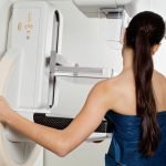
Age-related macular degeneration (AMD) is the leading cause of functional blindness in humans older than age 65 in developed countries around the world (American Academy of Ophthalmology, 2001). It is estimated that currently, 1.75 million people have progressive exudative (wet) AMD, and an additional 15 million have the less-progressive nonexudative (dry) form of AMD in the U.S. (Friedman et al., 2004; Mitchell et al., 2002; Klein et al., 1992). The incidence of wet AMD is expected to increase to 2.95 million affected by the year 2020 (Friedman et al., 2004). Incidence of AMD increases with age, from 10% in the decade spanning 65 to 75 years to 30% in the 75- to 85-year age group (Friedman et al., 2004; Mitchell et al., 2002; Klein et al., 1992). As the population ages, this condition will be a common presentation in physicians’ practices. NDs are in an excellent position to aid this group of patients through both prevention and treatment.
Pathogenesis
Known contributing factors to the pathogenesis of AMD include:
- Oxidative damage to the retinal pigment epithelium (RPE) and photoreceptors, which is exacerbated by low-quantity or dysfunctional macular pigments (lutein and zeaxanthin).
- Lipofuscin accumulation in the RPE.
- Accumulation of abnormal deposits in Bruch’s membrane, with resultant negative alterations in nutrient delivery and waste removal.Immunologic dysfunction at the Bruch’s – RPE interface (AAO, 2001).
Risk factors fall into known, potential and genetic categories. Known factors include smoking and age. Potential risk factors appear to be cardiovascular disease in general, hypertension, cataract formation and hyperopia (Klein et al., 2003; van Leeuwen et al., 2003; Ikram et al., 2003). Research supports the importance of genetic factors in AMD. (Meyers et al., 1995).
Diagnosis
AMD is currently defined as the presence of one or more of the following findings, without other etiology (AAO, 2001):
- Drusen formation
- Abnormalities (pigment changes) in the RPE
- Geographic atrophy of the RPE and choriocapillaris
- Neovascular or exudative maculopathy.
The basic difference between the two presentations of AMD is the presence or absence of choroidal neovascularization (CNV).
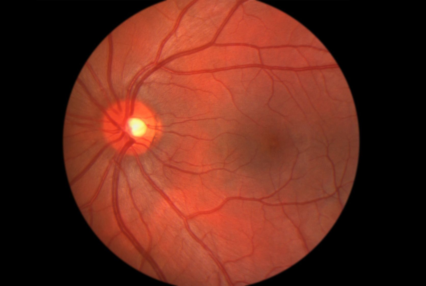
The dry form is hallmarked by the presence of drusen (yellow subretinal deposits), and often has gradual vision loss. Although the dry form is less threatening to vision, the drusen can become a point of architectural instability for the RPE and potentially lead to RPE detachment (Bradford, 2004).
In the wet form, progression of vision deficit is typically more rapid, and often the patient will present with “sudden” vision changes. These changes normally are not sudden, but the perception is often so due to the rapidly progressing nature of wet AMD. The CNV component of the wet form of AMD provides the major impetus for the rapid progression of vision loss. The full pathogenesis of CNV is not understood, but it is known at least to be a neovascularization/angiogenic phenomenon. The stimulus of this angiogenesis appears to be due at least in part to vascular endothelial growth factor (VEGF) and fibroblast growth factor (FGF).
Current Medical Treatment
The dry form of AMD is currently approached with one main treatment option: nutrient therapy. Nutrient approaches will be discussed, but it is important to remember that the eye physician community often recommends some form of macular supplement with at least vitamins C, E and beta-carotene with the minerals zinc and copper. It is the prevention model currently employed in the medical community for AMD.
In the wet form of the disease, treatment expands to focus on the CNV process. Options include anti-angiogenic medications, anti-inflammatory medications, laser photocoagulation and photodynamic therapy (PDT). Laser therapy is used to coagulate areas that are leaking and likely to cause greater, more rapid damage to vision. It is normally reserved for extrafoveal areas, as the negative aspect of laser is the obliteration of the retina in areas treated. It also is limited due to a high rate of recurrent CNV after treatment. The recurrence is to be expected, as the CNV is not addressed by the laser; it is simply employed to stop large leakage in the choriocapillaris. PDT is a relatively new laser treatment, and currently the only FDA-approved therapy for subfoveal CNV. A photosensitizing dye (verteporfin), which is taken up by the CNV, is activated by a 698nm nonthermal laser light. Platelet activation and thrombosis of the CNV occur without destruction of the overlying retinal receptor tissue (Bressler, 2001). Two anti-VEGF agents approved by the FDA are pegaptanib and ranibizumab. These agents show great promise, but require frequent (every four or six weeks) intravitreal injection as a delivery method. A steroid derivative angiostatic drug in phase III trials (anecortave acetate) has a six-month dose interval, and is delivered through a special sub-macular cannula.
In reading current ophthalmology literature, one is often impressed by the hope for improved treatment outcomes with current and emerging therapies. A closer look at the state of the art, however, leaves a less hopeful feeling when the costs, side effects and outcomes are considered. AMD of either form is, like all disease, best prevented or, at least, stopped from progression. This is certainly a tall order for medicine, and researchers are in pursuit of that goal. However, this next section outlines some therapies within the realm of naturopathic medicine that I believe comprise an improved therapeutic approach in the prevention and abatement of this disease process.
Naturopathic Therapies
As NDs are trained in and aware of aspects of safety and dosing where nutritional supplements are concerned, the following recommendations will not include a great deal of information regarding safety and dosing issues. Before employing any therapy, the physician should consider the entire patient and any co-morbid conditions.
Common nutrient combinations given by physicians outside the naturopathic arena in AMD follow the AREDS study formulation fairly closely, which includes:
500mg vitamin C, 400mg vitamin E, 15mg beta-carotene, 80mg of zinc as zinc oxide and 2mg of copper as cupric oxide (AREDS, 2001). In practice, these supplements appear to be of benefit to one degree or another.
Two categories of patients with AMD will present to NDs. Commonly, NDs will see those who are not laser or PDT candidates; and this nutrient combination with close monitoring is the therapy offered. Many patients present stating that they have been on these (AREDS) nutrients and their AMD is still progressing. Their options are limited in ophthalmology until they are candidates for laser or PDT.
The other category of patients is those who are post-laser or -PDT – or who have been told that these are the only other options available to them.
Either group can benefit from naturopathic intervention. Any specific therapy mentioned in this article is obviously best employed with a naturopathic approach to whole patient health. AMD patients, like all patients, need the basic determinants of health addressed. Prevention (whether prevention of AMD or of its progression) should be the first goal. Diet and nutrition optimization, blood sugar control, endocrine balance, digestive health and exercise should be foundational goals in these patients’ care.
When patients present with the complaint that they have been taking the standard AREDS-type supplements and are still progressing in their disease, they provide an excellent opening to discuss the determinants of health. Beyond that, it is helpful to point out that the AREDS supplementation is helpful but limited in its ability to help their disease process. AREDS study data seem to point to a 19%-25% drop in risk of progression of AMD in those patients at high risk of progression. It did not appear to be helpful for those at low risk or for those without disease.
Additional Support at Any Stage of AMD
Lutein and zeaxanthin, xanthophyll carotenoids, have an affinity for deposition in the fovea and macular tissue. They provide protection against blue and near-ultraviolet radiation for these sensitive macular tissues, as well as support antioxidant activity (Gale et al., 2003). Researchers believe that the macular pigment is entirely of dietary origin, and that both the healthy and diseased macula can accumulate and stabilize these pigments (Bone et al., 1997). These two compounds are found in corn, egg yolks and green vegetables and fruits, such as broccoli, green beans, green peas, brussel sprouts, cabbage, kale, collard greens, spinach, lettuce, kiwi and honeydew. Lutein and zeaxanthin are also found in nettles, algae and the petals of many yellow flowers (marigold is a common supplement source). Two concepts to consider in addition to supplementing these carotenoids: 1) they are sustained in the macula (and spared from degradation) by circulating antioxidants; and 2) in the diet, they compete for absorption with other carotenoids. Effective dose strengths are not agreed upon in oral administration, but a minimum of 20-60mg lutein and 5-10mg zeaxanthin daily are likely required in active disease.
Lycopene is also essential in degenerative eye conditions. It is found mainly in the ciliary body and RPE eye tissues. In addition to tomatoes (Lycopersicon esculentum) and tomato-based products – such as ketchup, pizza sauce, tomato juice and tomato paste (Gartner et al., 1997), lycopene is also found in watermelon, papaya, pink grapefruit and pink guava. Processed tomato products are more available dietary sources of lycopene than fresh tomatoes (Clinton et al., 1996, 1998). The average daily intake of lycopene is approximately 25mg in the U.S. Supplemental dosages in active macular disease should be at least 20mg to 100mg daily.
Other supplements to consider in the patient with AMD include omega 3 oils, coenzyme Q10, L-carnitine, vitamin C (as well as other glutathione support, such as NAC, alpha-lipoic acid and taurine) and the B-complex vitamins. Proper mitochondrial function, quenching of reactive oxygen species and support of glutathione function are reasonable support for patients dealing with a degenerative process (Richer, 2000).
Intravenous nutrient therapy is, in my experience, an excellent method through which tissue-specific nutrients can be delivered to the bloodstream and thus to the diseased areas, as well as to the body as a whole. It is often helpful in cases where long-term dietary and lifestyle inadequacy has left the patient with a system in need of much more than oral supplementation can deliver. It helps slow progression in many cases; raises nutrient tissue levels; and often allows diet, lifestyle and supplemental approaches to work more quickly and effectively. Of course, macular tissue that has undergone irreversible damage will not “see” again, but tissue in the process can slow or stop degenerative damage. See the accompanying case study for one approach to IV therapy in the AMD patient.
Age-related macular disease is an increasing malady of the visual health and quality of life for people age 65+ the world over. While medical therapy is progressing, and much effort is being expended in the research community on new therapies, a cure seems a long way off. In addition to this, current and emerging therapies carry significant cost, risks and, often, poor long-term outcomes. Naturopathic interventions that improve diet, lifestyle and overall health are still the best preventive measures available. In addition to addressing the determinants of health, the ND is in an excellent position to offer added therapies to help the patient with macular disease.
Case Study
An 83-year-old male with a diagnosis of wet AMD presented to my office with chronic complaint of vision loss secondary to advanced wet AMD. The patient had central vision deficit, which had been progressing for the past 10 years. He followed this progression with ophthalmologic service at a local teaching hospital. The patient wanted to look into his options with alternative therapy.
History:
- Progressive CV deficit X 10 years
- Loss of driving ability X 6 years
- Able to ambulate with care
- PMH includes CVD and CHF with CABG 6 years ago (Intermittent claudication in legs; hypertension, treated, with recalcitrant wide pulse pressure)
- Meds: Beta-blocker, diuretic, anti-arrhythmic; no vitamins/supplements
- Diet: Standard American diet (very low intake of any vegetable products).
PE:
- Ambulatory 83-year-old male in NAD
- EENT:
- Head and neck WNL
- Ears: Clear canals and TMs WNL
- Nose/throat: Clear, with no evidence of erythema, lymphadenopathy, discoloration or abnormal discharge.
- Eyes: Sclera BVI Gr2; media clear; no relative pupillary defect noted/PERRLA, Fundi revealed a normal optic nerve, C/D ratio of 0.3 OU, diffuse fundal geographic atrophy, occasional cotton-wool spots, and advanced macular scarring.
- Vision: Large central scotoma within 0-36 inches (unable to effectively use Amsler Grid Test). Intact peripheral fields by confrontation. Vision at 10 feet subjectively reveals patient able to “see that a picture frame is on the wall but not what the picture inside the frame is.”
Assessment:
- Progressively deteriorating. Wet AMD, followed by the ophthalmology service at 6-12-month intervals.
- Co-morbidity of advanced CV system disease
- Vision loss has significant negative effects on his ADL.
Plan:
- Diet advice given
- Full PARQ conference regarding the macular degeneration IV protocol.
- Patient consents to Tx to include six of the following IV treatments:
IV Formula:
Cr 600mcg
Vit C-500 50cc
Bicarb (8.4) 25cc
MgSO4 2g (4cc of 500mg)
Cal Gluc 5cc
KCL (40mEq) 3cc
Taurine 200mg
Carnitine 50mg
Infused in 500cc ½ NS over 1½-2 hours.
(Note: This is an older protocol. Currently, I will use the same formula with a decrease to 25cc C-500 and addition of a 1g glutathione push at the end of the IV.)
First treatment:
- Patient tolerates IV well, with no significant change in pre-post Tx vitals
- At the end of the IV, he notes that there is a “deer in that picture on the wall.”
- He is excited to return.
Second treatment:
- Patient in for IV #2 (weekly interval).
- No problems noted after last Tx.
- Vitals are stable/no venous sn/sx of concern present
- OK to administer IV #2
- Patient reports vision change based on the picture on the wall: Back to no picture prior to IV treatment and some picture after IV.
Sixth treatment:
- Patient has mild retention of some central vision.
- Sees some of the picture at the start of the IV, and more at the end.
- Is very pleased.
- Is off to see the ophthalmologist in 60 days and will report back.
Follow-up:
- Saw ophthalmologist
- Vision and fundus report: No progression of vision loss or fundus pathology. This is the first time since this patient has been followed by this service that there has been no advance in the pathology noted.
As a note, the patient never told his ophthalmologist about the IV therapy or the follow-up nutritional therapy. The ophthalmologist was impressed enough with the disease abatement (and probably knew he had done something) that she told him, “If you think of anything you have changed in the last six months, please keep doing it.”
Plan for ongoing Tx and F/U
- Slow increase in water intake
- Increase dark greens
Rx:
- A whole food supplement supplying significant Brassicaceae and other eye-specific nutrients.
- Multivitamin, EPA/DHA, plus the ARED formula
- F/U PRN.
I did not see this patient again, but was able to follow up through his daughter. He was pleased with the abatement in his visual loss and disease progression, as was his ophthalmologist. After two years, he reported no advancement in vision loss.

Paul Anderson, ND is a naturopathic physician practicing in Seattle, Wash., and Tempe, Ariz. He is a graduate of NCNM, and a full-time core faculty member at Bastyr University, teaching in the clinical science division.
References
American Academy of Ophthalmology Retina Panel: Age Related Macular Degeneration, Preferred Practice Pattern, San Francisco, 2001, American Academy of Ophthalmology.
Friedman DS et al: Prevalence of age-related macular degeneration in the United States, Arch Ophthalmol Apr;122(4):564-72, 2004.
Mitchell P et al: Five-year incidence of age-related maculopathy lesions: the blue mountains eye study, Ophthalmology June;109(6):1092-7, 2002.
Klein R et al: Prevalence of age-related maculopathy. the beaver dam eye study, Ophthalmology June;99(6):933-43, 1992.
Klein R et al: The five-year incidence and progression of age-related maculopathy. the beaver dam eye study, Ophthalmology Jan;104(1):7-21, 1997.
Vingerling JR et al: AMD and smoking. the rotterdam study, Arch Ophthalmol Oct; 114(10):1193-6, 1996.
Klein R et al: The association of cardiovascular disease with long-term incidence of age-related maculopathy: the beaver dam eye study, Ophthalmology June;110(6):1273-80, 2003.
Van Leeuwen R et al: Blood pressure, atherosclerosis, and the incidence of age-related maculopathy: the rotterdam study, Invest Ophthalmol Vis Sci Sep;44(9):3771-7, 2003.
Ikram MK et al: Relationship between refraction and prevalent as well as incident age-related maculopathy: the rotterdam study, Invest Ophthalmol Vis Sci Sep;44(9):3778-82, 2003.
Meyers SM et al: A twin study of age-related macular degeneration, Am J Ophthalmol Dec;120(6):757-66, 1995.
Bradford CA: Basic Ophthalmology (8th ed), San Francisco, 2004, American Academy of Ophthalmology.
Bressler NM: Photodynamic therapy of subfoveal choroidal neovascularization in age-related macular degeneration with verteporfin: two-year results of 2 randomized clinical trials-tap report 2, Arch Ophthalmol Feb;119(2):198-207, 2001.
Age-Related Eye Disease Study Research Group: A randomized placebo-controlled, clinical trial of high-dose supplementation with vitamins C and E, beta-carotene and zinc for age-related macular degeneration and vision loss: AREDS report no. 8, Arch Ophthalmol Oct;119(10):1417-36, 2001.
Fletcher RH, Fairfield KM: Vitamins for chronic disease prevention in adults; clinical applications, JAMA Jun19;287(23):3127-9, 2002.
Gale CR et al: Lutein and zeaxanthin status and risk of age-related macular degeneration, Invest Ophthalmol Vis Sci Jun;44(6):2461-5, 2003.
Bone RA et al: Distribution of lutein and zeaxanthin stereoisomers in the human retina, Exp Eye Res Feb;64(2):211-8, 1997.
Gartner C et al: Lycopene is more bioavailable from tomato paste than from fresh tomatoes, Am J Clin Nutr.66:116-122, 1997.
Clinton SK: Lycopene: chemistry, biology, and implications for human health and disease, Nutr Rev. 56:35-51, 1998.
Clinton SK et al: Cis-trans lycopene isomers, carotenoids, and retinol in the human prostate, Cancer Epidemiol Biomarkers Prev. 5:823-833, 1996.
Giovannucci E: Tomatoes, tomato-based products, lycopene, and cancer: review of the epidemiologic literature, J Natl Cancer Inst. 91:317-331, 1999.
Bone RA et al: Lutein and zeaxanthin in the eyes, serum and diet of human subjects, Exp Eye Res. 71:239-245, 2000.
Kostic D et al: Intestinal absorption, serum clearance, and interactions between lutein and beta-carotene when administered to human adults in separate or combined oral doses, Am J Clin Nutr. 62:604-610, 1995.
Landrum JT et al: A one-year study of the macular pigment: the effect of 140 days of a lutein supplement, Exp Eye Res. 65:57-62, 1997.
Richer S: Antioxidants and the eye, Int Ophthalmol Clin 40(44)1-16; Fall 2000.


