

Unique Imaging Technique Used for Determining Cellular Progression of Diabetic Retinopathy
Node Smith, ND
A team of researchers from University of Wisconsin-Milwaukee has developed an imaging method that shows what is happening on a cellular level in the human retina when it is exposed to prolonged hyperglycemic states.1 Currently, there is no imaging technique that allows visualization of biochemical changes occurring in the retinas of diabetic patients.
Diabetic retinopathy
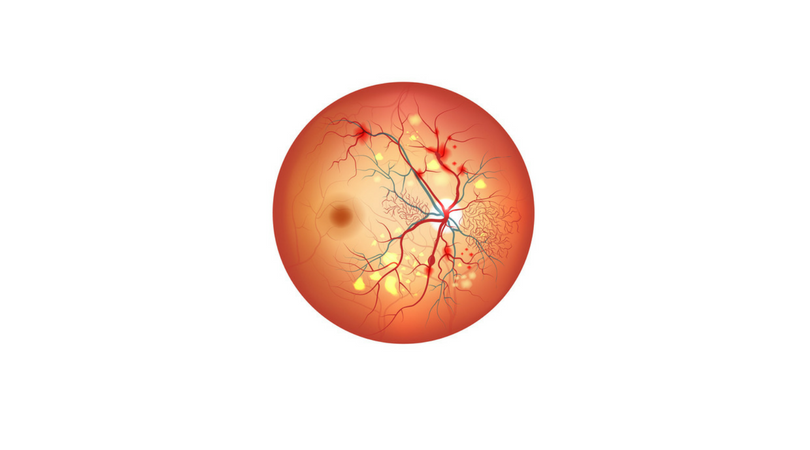
The retina is a sensitive aspect of the eye, which is responsible for converting light signals into neural signals that the brain interprets as vision. The retina is a commonly damaged end organ in diabetes, and such damage results in diabetic retinopathy, a major cause of blindness.
Changes within the retinal cells drive the disease
The team of researchers from UWM has shown these changes within the retinal cells that actually drive the disease. They utilized a unique imaging technique that allowed for identification of biomarkers significant for cellular damage. The imaging tool concentrates the light of a synchrotron in infrared wavelengths, while using Fourier transform infrared imaging (FT-IR) to produce a molecular blueprint of various biomarkers in the cells in question.
Fourier transform infrared imaging (FT-IR)
It was seen that the photoreceptors in the retina are the most susceptible to damage at the beginning of disease progression, and 6 weeks into disease, only the retinal layer showed damage. The other layers appeared healthy. This confirms earlier research that has suggested an irregular retinal deterioration. What’s new is that this imaging technique was able to show that significant cellular damage is occurring in the first 3 months.
Disease progression noted in the studies
The disease progression noted in the studies showed a steady rate of cellular deterioration from 6 – 12 weeks of disease, followed by a decrease between 3 – 6 months. Disease progression then gradually increased by month 10 of disease. The greatest amount of cellular damage was noted at 6 months into disease. The variations in disease progression is thought to follow cellular mechanisms of repair that are being overwhelmed at different rates by the damaged and degraded proteins.
The imaging platform used to visualize the damaged cellular components may be a platform for studying other diseases of the body.
Source:
- Aboualizadeh E, Hirschmugl CJ. Highlighting IR Spectrochemical Imaging of the Retina. Trends Biochem Sci. 2018.
Image Copyright: <a href=’https://www.123rf.com/profile_guniita’>guniita / 123RF Stock Photo</a>
 Node Smith, ND, is a naturopathic physician in Portland, OR and associate editor for NDNR. He has been instrumental in maintaining a firm connection to the philosophy and heritage of naturopathic medicine among the next generation of docs. He helped found the first multi-generational experiential retreat, which brings elders, alumni, and students together for a weekend camp-out where naturopathic medicine and medical philosophy are experienced in nature. Four years ago he helped found the non-profit, Association for Naturopathic ReVitalization (ANR), for which he serves as the board chairman. ANR has a mission to inspire health practitioners to embody the naturopathic principles through experiential education. Node also has a firm belief that the next era of naturopathic medicine will see a resurgence of in-patient facilities which use fasting, earthing, hydrotherapy and homeopathy to bring people back from chronic diseases of modern living; he is involved in numerous conversations and projects to bring about this vision.


