

Diagnostic Imaging for Musculoskeletal Concerns
Sam Russo, ND
Diagnostic imaging is an integral part of musculoskeletal medicine practice. The last century saw an incredible development in the field of diagnostic imaging, from the refinement of radiography to the advent of various types of magnetic resonance imaging (MRI) and positron-emission tomography (PET) scans. However, I believe that no imaging modality is as safe, accessible and affordable as ultrasound.
Ultrasound imaging has been in use since the 1950s. In the last 30 years, ultrasound has developed into an indispensable tool for practitioners specializing in musculoskeletal medicine. In just the last few years, the refinement of high-frequency broadband transducers has provided a range of 4-18 MHz, allowing deep tissue penetration with low frequency and high-resolution imaging with high-frequency probes. Compounded imaging and speckle reduction technology have allowed for amazingly detailed examinations. Sensitive color and power Doppler technology have improved the ability to evaluate for vascularity and hyperemia in inflamed tissues, including connective tissue structures as well as peripheral nerves.
Ultrasound can be used to assess muscular atrophy, adhesive capsulitis, joint effusions, bursitis, tendinosis, calcific tendonitis, osteoarthritis of joint margins, hyaline cartilage thickness and defects, ligament injury and nerve entrapments. Neuromusculoskeletal structures amenable to evaluation with ultrasound include all of the extremities, the sacrum and external pelvis.
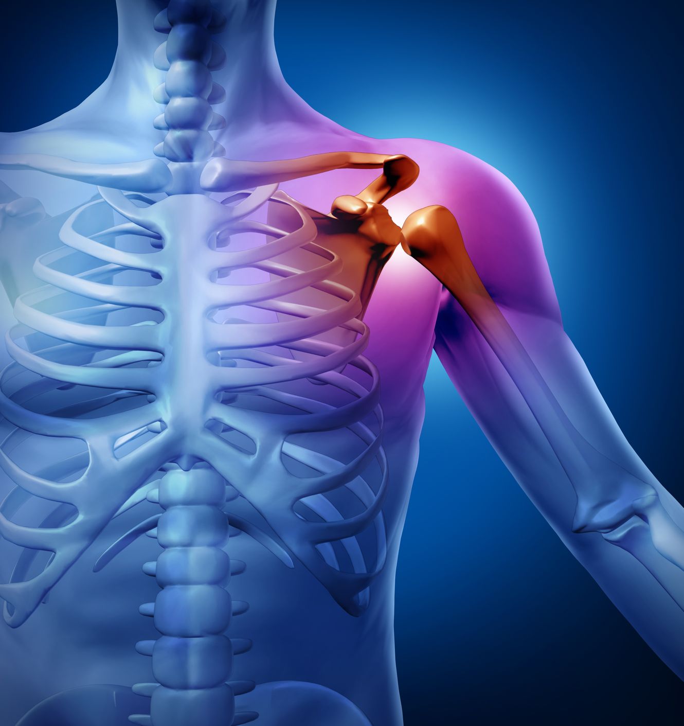
Ultrasound Examination of the Shoulder
Rotator Cuff Lesions
Ultrasound is a valuable tool for the evaluation of rotator cuff pathology. In a study to determine the effect of the one-stop clinic on the interval and delay in management of patients with suspected rotator cuff pathology, 74 patients were evaluated with ultrasound, then reviewed retrospectively. Ultrasound had an overall sensitivity of 93.75%, specificity of 100% and accuracy of 91% for full-thickness tears. In-office use of ultrasound also decreased the time to development of a treatment plan (Miller et al., 2008). In a similar study, ultrasound evaluation of the rotator cuff was able to predict treatment outcome of patients with full-thickness tears, who were more likely to require surgical intervention compared to patients with partial tears, who generally had improvement with conservative measures despite patient demographics, clinical conditions and physical examination findings (Ferrari et al., 2002). Of note, MRI is not a sensitive tool when assessing partial thickness tears, although magnetic resonance (MR) arthrography is effective in assessing articular-sided lesions, but is also an invasive procedure. Two recent studies demonstrated the sensitivity of MR arthrography to be 84%-95% and the specificity to be 96%-100% for partial-thickness articular surface tears of the rotator cuff (Ferrari et al., 2002; Meister et al., 2004). With that consideration, however, a study of 282 shoulder sonograms in patients ultimately treated surgically demonstrated a sensitivity, specificity, positive predictive value and negative predictive value of 94.1%, 96.1%, 96.6% and 93.2%, respectively, for partial-thickness tears (Ziegler, 2004).
Shoulder Instability
A Hill-Sachs lesion is a posterolateral defect in the humeral head. It is a definitive indication of previous anterior shoulder subluxation or dislocation, and a clear indicator of the direction of shoulder instability. It is also very difficult to evaluate with most imaging techniques, and the gold standard of diagnosis is a surgical finding. However, in a study of 61 patients with anterior shoulder dislocation, 52 were found to have a Hill-Sachs lesion on sonography vs. 54 on surgical exploration, making ultrasound 96% sensitive and 100% specific (7 of 7 negative cases), with a diagnostic accuracy of 97% (Cicak et al., 1998). Ultrasound is also a complementary imaging technique for the evaluation of posterior shoulder instability and dislocation, which are easier to misdiagnose with conventional radiographs. Ultrasound is not effective in the assessment of the superior glenoid labrum, however.
Ultrasound Exam of Nerve Entrapments
Most common nerve entrapments that occur in osteofibrous tunnels are amenable to ultrasound examination. In the upper limb, these tunnels include the carpal tunnel for the median nerve, and the cubital and Guyon tunnels for the ulnar nerve. In the lower limb, they include the fibular neck for the common peroneal nerve, the tarsal tunnel for the posterior tibial nerve and the intermetatarsal spaces for the interdigital nerves.
- Carpal Tunnel Syndrome (CTS): The ultrasonographic measurements of the cross-sectional area of the median nerve are increased significantly in patients with CTS when compared with controls in many studies. Ultrasound is comparable to electrodiagnostic studies for the diagnosis of CTS and is not an invasive procedure (Wong et al., 2004).
- Tarsal Tunnel Syndrome: Tarsal tunnel syndrome is a neuropathic entrapment of the tibial nerve and its branches on the medial side of the ankle, and is characterized by paresthesia and pain in the toes and planta pedis. Evaluation of 17 patients with tarsal tunnel syndrome with preoperative ultrasonography showed 100% consistency with surgical findings of common causes of tarsal tunnel syndrome, including ganglia, talocalcaneal coalition (progressive loss of movement of mid-foot joints occurring due to a spontaneous union) and varicose veins, which can occur singly or in combination (Nagaoka and Matsuzaki, 2005). Other causes of tarsal tunnel syndrome that can be evaluated with ultrasound include post-traumatic fibrosis, tenosynovitis, and tumor of the nerves or other structures.
Ultrasound-Guided Interventions
Ultrasound guidance has also been used for aspiration and injection of the joints of the extremities as well as the spine, ganglion cysts, bursae and nerve blocks.
Diagnostic injections can be used to successfully differentiate a wide variety of conditions. For example, diagnostic injections can differentiate the cause of pain in many disorders, including osteoarthritis, rheumatoid arthritis, labral tears and capsulitis.
Intra-articular injection of the hip is a good example of how ultrasound guidance improves accuracy. Needles placed using only surface landmarks (“blind”) often miss the joint entirely. Blind injections accurately enter the hip joint only 52%-80% of the time and may pass within 4.5 mm of the femoral nerve. In contrast, the accuracy of sonographically placed needles for intra-articular hip injections was 97% as confirmed by fluoroscopy for 30 injections in an outpatient pain management study (Smith et al., 2009). In the shoulder, the accuracy for blind intra-articular injection of the glenohumeral joint ranges from 26.8%-80% for an anterior approach and is about 50% from a posterior approach (Sethi et al., 2005; Sethi and El Attrache, 2006). A successful injection occurred more often with ultrasound (94%) than with fluoroscopic guidance (72%) in both anterior and posterior approaches in a comparison study (Rutten et al., 2009).
Ultrasound vs. Other Imaging Techniques
An ultrasound exam occurs in an outpatient office setting whereas an MRI or MR arthrography requires a visit to a hospital or radiologist. Some sonographic units are even portable. Sonography is equitable in cost to radiographs, and costs about one-sixth the price of a CT scan and one-tenth the price of an MRI. The resolution of ultrasound is comparable and can exceed that of MRI. Ultrasound also allows real-time or dynamic evaluation of joint tendons and can be used as a guide for percutaneous procedures. Unlike MRI, ultrasound can evaluate joints containing orthopedic hardware.
Limitations of ultrasound include a smaller field of view and limited visualization of subcortical bone and bone marrow abnormalities. For example, MRI has the ability to fully evaluate anatomic areas that cannot be accessed by ultrasound – including bone marrow and areas of joint cartilage deep within a joint – unless the surface can be exposed to the ultrasound transducer. One major advantage of other imaging modalities over ultrasound is the lack of operator dependence; quality imaging is achieved through standard protocols. Currently, the quality of ultrasound is operator dependent, although protocols are being developed to standardize the views and dynamic tests for examination of each area of the upper and lower extremity.
Case Study
A 50-year-old woman was referred for evaluation and recommendations for treatment of left shoulder pain after a hiking accident where she fell on the side of her extended left arm. She had no pain with her arm hanging at her side. She had restricted abduction and flexion with pain about midway to her shoulder. She could flex her elbow and internally rotate her shoulder with her arm at her side. She could have pain at night if she got into a wrong position. She had not had any shoulder problem like this before. She is left handed. She had not had any imaging of her left shoulder, either before or after her fall.
Physical exam: DTR +2 for triceps; biceps +1, brachioradialis +1 bilaterally. Sense grossly intact in feet. Examination of the left shoulder and arm demonstrated circulation grossly intact in left hand. No sulcus sign. Full passive abduction and flexion. Active motion limited to 80 deg of flexion, 65 deg of abduction, both of which were painful midway through this range. 3/5 strength in supraspinatus and empty can test without pain. Positive drop arm test. The patient had some pain with external rotation with arm at her side, which increased if she abducted to 45 degrees then externally rotated. She had no pain with active extension. Biceps testing was supination pain free but weak, 3/5.
Ultrasound examination of the right shoulder demonstrated an intact biceps tendon well seated in the bicipital groove that did not sublux with dynamic testing. On transverse imaging, a halo was present around the biceps tendon, indicating fluid within the biceps tendon sheath. There was increased flow on color flow mapping (CFM) viewing, indicative of an inflammatory process (see Figure 1). Dynamic testing of the subacromial space showed subacromial impingement flexion (subacromial space=0.54 cm, N>.7 cm). There was an articular surface defect in the supraspinatus tendon measuring 0.9 cm and a 0.3 cm deep full-thickness tear as it communicates with the subdeltoid bursa, which was enlarged at 0.3 cm thick over the defect. This defect was present on both transverse and longitudinal views, and may extend to the articular surface of the infraspinatus (see Figure 2). The superior aspect of the posterior labrum had an anechoic cleft, which may indicate detachment or a labral tear (see Figure 3). Unremarkable structures: subscapularis tendon, infraspinatus tendon, acromioclavicular joint.
 Sam Russo, ND has a practice in musculoskeletal medicine in Winooski, Vt. He is a member of the Naturopathic Academy of Therapeutic Injection, American Academy of Pain Management, Vermont Association of Naturopathic Physicians, Vermont Association of Acupuncture and Oriental Medicine (past president), American Institute for Ultrasound in Medicine and American Association of Naturopathic Physicians. Dr. Russo currently serves as an advisor to the Office of Professional Regulation in Vermont.
References
Bianchi S et al: Sonographic evaluation of posterior instability and dislocation of the shoulder: prospective study, J Ultrasound Med 13:389-393, 1994.
Cicak N et al: Hill-Sachs lesion in recurrent shoulder dislocation: sonographic detection, J Ultrasound Med 17:557-560, 1998.
Ferrari FS et: Supraspinatus tendon tears: comparison of US and MR arthrography with surgical correlation, Eur Radiol 2002;12(5):1211–17.
Meister K et al: MR arthrography of partial thickness tears of the undersurface of the rotator cuff: an arthroscopic correlation, Skeletal Radiol Mar;33(3):136-41, 2004.
Miller D et al: A “one-stop clinic” for the diagnosis and management of rotator cuff pathology: getting the right diagnosis first time, Int J Clin Pract 62(5):750-753, 2008.
Nagaoka M, Matsuzaki H: Ultrasonography in tarsal tunnel syndrome, J Ultrasound Med 24:1035-1040, 2005.
Rutten M et al: Glenohumeral joint injection: a comparative study of ultrasound and fluoroscopically guided techniques before MR arthrography, Eur Rad 19(3):722-730, 2009.
Sethi P, El Attrache N: Accuracy of intra-articular injection of the glenohumeral joint: a cadaveric study, Orthopedics 29:149-52, 2006.
Sethi P et al: Accuracy of anterior intra-articular injection of the glenohumeral joint, Arthroscopy: The Journal of Arthroscopic & Related Surgery 21(1): 77-80, 2005.
Smith J et al: Accuracy of sonographically guided intra-articular injections in the native adult hip, J Ultrasound Med 28:329-335, 2009.
Wong SM et al: Carpal tunnel syndrome: diagnostic usefulness of sonography, Radiology 232:93-99, 2004.
Ziegler DW: The use of in-office, orthopaedist-performed ultrasound of the shoulder to evaluate and manage rotator cuff disorders, J Shoulder Elbow Surg May-Jun;13(3):291-7, 2004.


