

RIT of the Knee: Case No. 1
David A. Tallman, DC, NMD
Regenerative injection therapy (RIT) applied as part of a comprehensive naturopathic orthopedic program is an extremely effective approach for the treatment of osteoarthrosis of the knee. When a diagnosis is achieved which logically accounts for the present pathophysiology and symptomology of the affected joint, a physician carefully administering RIT will alter the course of the patient’s symptoms. This is to not only reduce the pain and dysfunction of osteoarthrosis (OA) or degenerative joint disease (DJD), but will set the stage for cellular regeneration that is commensurate with a pain-free, biomechanically correct articulation. This case example demonstrates the inherent power of the body’s recuperative abilities upon applying precision RIT for chronic OA of the knee.
Rehabilitation, detoxification and dietary measures accompanied the treatment program to facilitate optimum tissue anabolism, and I find that applying the naturopathic paradigm (diet, detoxification, exercise and rest) greatly hastens the patient’s treatment goals (especially when your patients are compliant).
A review of the published literature concerning RIT of the knee reveals promising outcomes. Reeves et al in 2003 concluded what was a three-year follow-up of a group of subjects that underwent multiple intra-articular dextrose injections for ACL laxity. A lasting benefit of normalizing ACL tightness, increasing pain-free range of motion (ROM), and decreasing daily pain levels was demonstrated.
In a paper published in 2000 by the same author, significant radiographic improvements of osteoarthritis of the knee were noted in the form of decreased osteophyte size of the femur and tibia. The authors also noted what appeared to be an increase in medial and lateral tibiofemoral compartment cartilage width in many of the subjects.
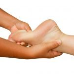
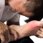
This patient was a 63-year-old female that presented with chronic bilateral knee discomfort that had an insidious onset over the past few years. She reported a history of playing tennis frequently throughout her life, but she could no longer play tennis without difficulty and aggravation of the current joint symptoms. She reported that going up and down stairs was difficult and that symptoms had slowly become more and more noticeable. Both knees were causing her daily discomfort, with the right being slightly worse than the left. She experienced exacerbation of pain with continuous joint demand such as walking long distances, dancing and exercising. The patient stated that many of her friends who have had surgery reported unfavorable outcomes, and she wished to avoid a surgical procedure.
Orthopedic Exam
The orthopedic exam began with the observance of antalgia upon arising from a seated position. The patient also appeared to have mild antalgic gait due to her knees. Unilateral deep knee bends produced discomfort bilaterally, and palpation of the joint while bending demonstrated crepitus, the right knee being worse. Valgus and varus stresses did not produce gross laxity but did produce tenderness in the medial compartment upon genu-valgus stress bilaterally, attributed pinching the soft tissue between the femoral condyle and the medial tibial plateau; namely, the degenerating medial meniscus and the femoral cartilage.
Palpation of the medial collateral ligament attachments on the femur and tibia were tender, as were the lateral collateral attachments on the fibular head and femur. The pes anserine attachments were also tender to light palpation bilaterally. Compression of both the medial and lateral meniscus revealed discomfort upon applying axial force against it bilaterally. Sitting palpation of the medial meniscus demonstrated increased tenderness upon internal rotation of the tibia, and the coronary ligament attachments were also very tender on the widened tibial plateau.
Radiographs were ordered prior to the treatment to assess the extent and pattern of the femoral condylar and tibial plateau osseous derangements, as well as to asses the relationship between the fibular head and the tibia. Weight-bearing AP and lateral views were taken. Radiographs offer invaluable information for achieving a working diagnosis by demonstrating trabecular line patterns that are not appreciable in other forms of imaging. Plain films are requisite before I administer RIT. Her radiographs demonstrated significant osteophytosis, sclerosis, periosteal changes and loss of cartilage height.
She was diagnosed with chronic sprains of the lateral collateral, medial collateral and coronary ligaments of the femur, tibia and fibular attachments. She also had mild laxity of the anterior cruciate ligaments, secondary myospasms around the knee joint, degeneration of articular cartilage, and menisci secondary to the chronis sprains.
Treatment
The medical plan was to restore the integrity of the ligaments, the articular capsule and the condylar and plateau linings. Regenerative injection therapy was recommended and would be commensurate with her response. Prolotherapy with needle manipulation (a custom technique I devised) was applied to the collateral and coronary ligaments, intra-articular prolotherapy was alternated with viscosupplementation with sodium hyaluronate. She was treated six times over a seven-month period. The patient was prescribed MSM and vegetarian glucosamine to be taken every four hours. She was advised to dose turmeric as needed for pain. Her pain scores improved with each four-week follow-up after each treatment.
At her six-month follow up, the patient was able to go up and down stairs without difficulty, and activities of daily living (ADL) were no longer painful. She told me she believed she was no longer a candidate for joint-replacement surgery. Another plain film was ordered to evaluate trabecular and articular changes that confirmed the positive results.
Discussion
What was of the highest importance in this case was the restoration of “physiologic biomechanics” versus her initial presenting of “pathologic biomechanics” caused by ligaments that have lost their integrity from the sudden lateral and medial shocks from chasing a tennis ball for many years. The quick changes to the body’s velocity place the ligaments in a compromised position. The roots of the ligaments’ boney attachment at the fibro-osseous junction need to be physiologically (or naturopathically) stable if the physician is to expect lasting results with the knee.
References
A systematic review of prolotherapy for chronic musculoskeletal pain. Clin J Sport Med. 2005 Sep;15(5):E376
David A. Tallman, DC, NMD, studied at the Ohio State University for his undergraduate education. He then attended Texas Chiropractic College in Pasadena, Texas, where he received a Doctorate of Chiropractic with an internship focus on orthopedics and radiology. He earned a Doctorate of Naturopathic Medicine at Southwest college of Naturopathic Medicine in Tempe, Arizona. Dr. Tallman is in private practice in Scottsdale, Arizona where he treats orthopedic and rheumatological conditions.

