

Mixed Inflammatory Skin Conditions: Emerging Dermatological Entities
Nadia Arora, ND
Clinical Case Studies
Coexisting skin disorders are a well-known phenomenon. For example, superimposed bacterial infections are often found in patients with eczema or psoriasis. In my practice, I am more frequently coming across the puzzling coexistence of several dermatological disorders in the same individual. Those were typically eczema and psoriasis. In the majority of such cases, I believe that psoriasis, an autoimmune skin condition with many faces, was misdiagnosed as eczema. Another chameleon, dermatitis herpetiformis, was often mistaken for either eczema or psoriasis. However, I have come across patients who had eczema and psoriasis or even eczema, psoriasis, and dermatitis herpetiformis simultaneously. These cases were confirmed histologically and had distinctly different presentation and different response to treatment. Three of these cases are described further herein.
Mixed Eczematous and Psoriatic Lesions in a Patient With Allergies
Linda is a 37-year-old African American female who was diagnosed as having eczema in her late teens. She has a history of environmental allergies and asthma. She was never tested for food allergies but believes that she has some because she noticed an increased number of lesions and increased pruritus after consuming certain foods. At the time she was seen in my office for the initial consultation, her eczema was widespread, covering most of her body, and was extremely itchy and painful. The lesions on the legs were very thick, and she reported such a tightening of her skin that it was making it difficult for her to move. She was treated with prednisone, both oral and topical, as well as cyclosporine topically. She reported no improvement of her lesions, except that oral prednisone was making the itching more tolerable. She was not using any topical treatments for her facial lesions. She was concerned about long-term effects of corticosteroid use and wanted to explore other options. One month before our visit, she had discontinued prednisone by titrating it slowly and had decided not to resume it.
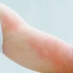
On physical examination, Linda’s skin was hot to touch and excessively dry. The lesions, which were covering most of her body, including her face and eyelids, appeared thick, dry, and scaly. The lesions on the extremities had the appearance of large sheets of silvery plaques, with multiple cracks in the plaques. In the cracks, the raw underlying tissue could be seen, as well as some dry blood.
I noted that Linda’s blood pressure was high (140/87 mm Hg) and that her heart rate was increased (102 beats/min). The patient reported that her blood pressure had increased over the past 2 months since she had started on cyclosporine and oral prednisone. There were inspiratory wheezes on auscultation, as well as a systolic heart murmur. The abdomen was tender in the periumbilical area, and there was moderate nontender splenomegaly. The thyroid was boggy and enlarged.
We discussed the possibility of her condition being mixed eczema and psoriasis, given the features. We ordered a complete blood cell count, vitamin D3 levels, a nonceliac gluten sensitivity panel, and thyrotropin level.
The patient was instructed to start on quercetin (1000 mg/d), fish oil (2000 mg/d), and vitamin D (5000 IU/d). Her test results came back positive for nonceliac gluten sensitivity, with native antigliadin antibodies at 68 U (normal, 0-19 U).Her vitamin D (25-hydroxyvitamin D) level was 14 nmol/L (normal, 34-100 nmol/L). Her thyrotropin level was 2.1 mIU/L, and her complete blood cell count showed microcytic anemia. Both anemia and vitamin D deficiency were, in my understanding, caused by malabsorption related to gluten sensitivity. Linda was advised to start a gluten-free diet as soon as possible.
I suspected that her anemia was most likely of a mixed type: iron deficiency and chronic disease. We decided not to supplement iron because it could worsen constipation. Instead, she was advised to increase iron-containing foods in her diet. We chose to increase vitamin D3 supplementation to 8000 IU/d for 2 months. In my experience, supplementation with vitamin D tends to increase blood levels of vitamin D faster. In addition, when blood levels are low and there is an inflammatory and uncomfortable skin condition, steady supplementation with high-dose vitamin D can provide additional relief, particularly in the late fall to winter months, when sun exposure is minimal.
Over the course of the next 3 months, I kept in touch with Linda. She briefly informed me that her skin had started to improve.
I saw her for a follow-up visit in February 2013. Linda was very pleased with the progress that she had achieved. She reported that her skin had started to improve steadily within approximately 1 month after removal of gluten from her diet. She reported that the discomfort and itching improved in a matter of days after she had started taking quercetin. In Linda’s words, “Quercetin works just as well as prednisone worked, when I first started, without side effects!” Her facial lesions had resolved almost completely. The lesions on her legs were thinning, and new skin was showing through. What is most important, these lesions did not feel as tight anymore; the cracks in the plaques had started healing, and she was able to move without pain.
Although Linda still had numerous lesions, particularly those on the back of her neck, which did not seem to reduce in size, a shift in her condition was remarkable. On an interesting note, the psoriatic lesions exhibited the most improvement. She was advised to continue on her current treatment plan. We retested her blood level of vitamin D; it was 39 nmol/L, a good improvement from 3 months ago. I recommended reduction of vitamin D3 supplementation to 3000 IU/d. I believe that it might be worthy to biopsy both types of Linda’s lesions to determine whether they are indeed different histologically, but this patient is not willing to undergo a biopsy at this time.
Eczema vs Psoriasis
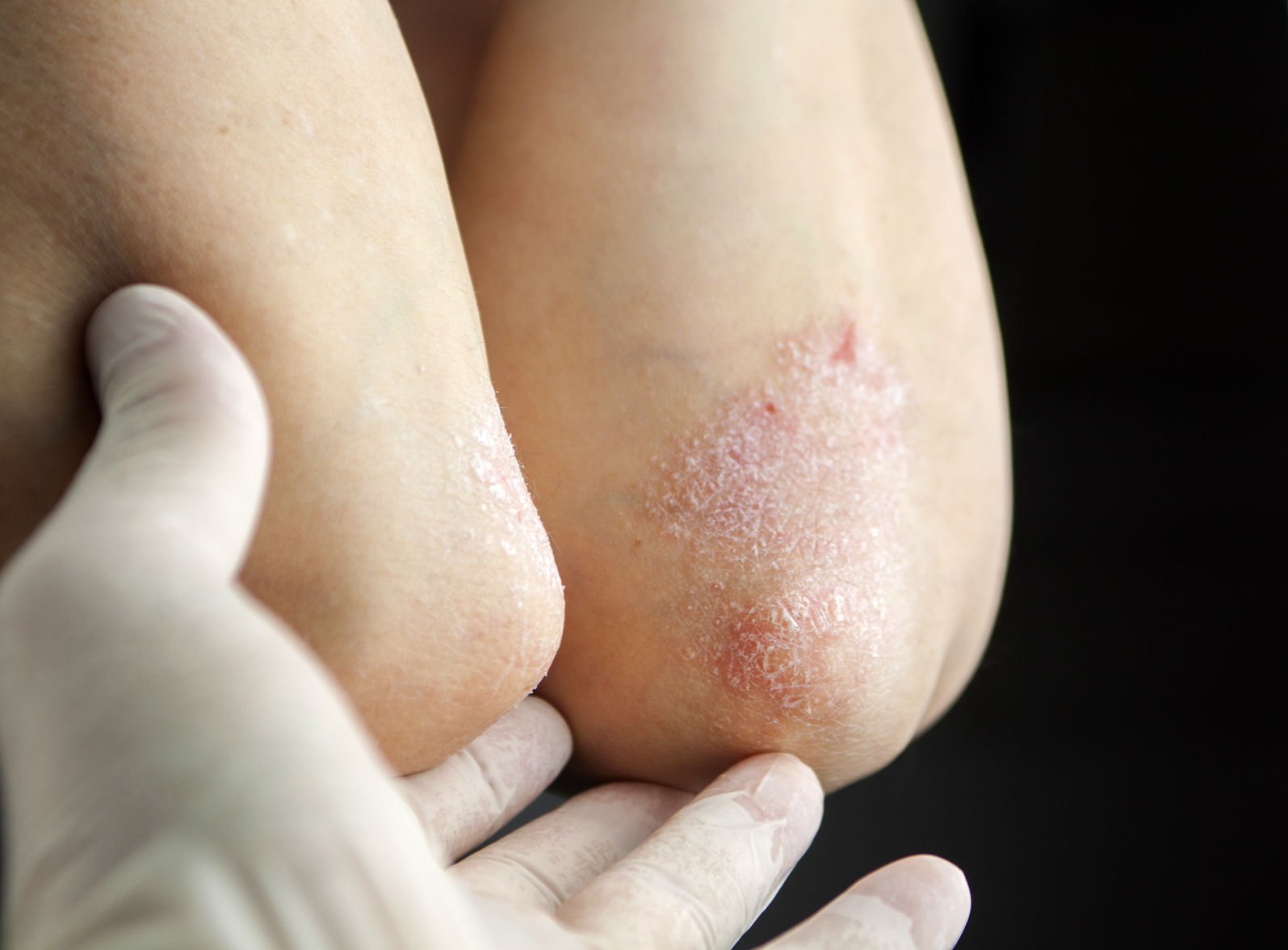
Theoretically, clinical differentiation between eczema and psoriasis should be easy because it is based on certain well-known characteristics of each. For example, eczema typically presents with dry, erythematous, inflammatory lesions of sandy texture and often prefers moist hidden areas of the body such as skin folds. On the other hand, classic psoriasis has thick, scaly, silvery plaques that are found on areas that are exposed to pressure and trauma such as elbows and knees. There is a challenge, though, and it comes with the fact that psoriasis is an entity with many faces. A plaque psoriasis, characterized by silvery, thick plaques, typically found on extensor surfaces, is easy to recognize. Other types of psoriasis such as pustular psoriasis, which typically affects the palms and the soles of the feet, or inverse psoriasis (dry inflammatory cracks found in skin folds) are often misdiagnosed as eczema. Again, the distinction could theoretically be made clinically in the process of observing the behavior of the lesions. Eczema typically gets worse in the summer, from increased sweating and humidity, whereas psoriasis improves in the summer, mainly due to exposure to sunlight and increased levels of vitamin D. In practice, lesions can behave differently from the predicted behavior. For example, eczema might improve from exposure to sunlight, and psoriasis (particularly inverse psoriasis) might worsen during the hot humid summer months.
The mechanism of improvement of eczema from exposure to sunlight is not fully understood. Emerging data on immunomodulating effects of sunlight suggest that UV light suppresses the skin immune response, an effect that is helpful in cases of exaggerated immune reaction of eczema and psoriasis.1 Simultaneously, UV light irradiation upregulates expression of skin antimicrobial proteins, thus preventing superimposed infections, common in eczema.2 A healing effect of the sun might be rendered by suppression of oxidative stress via activation of the p53 gene in skin, an effect that we are only beginning to explore.3 The strongest effect, however, is probably rendered by an increased production of vitamin D in skin.4,5 This is also supported by my observation that individuals with eczema or with mixed eczematous and psoriatic lesions tend to improve with the supplementation of high doses of vitamin D3, even in the absence of sufficient sunlight exposure.
Our next case is intended to illustrate an atypical behavior of eczema. It demonstrates how lesions that are morphologically eczematous can behave in a way that is typical of psoriasis.
Eczema, Misbehaving
Natasha, a 24-month-old white female, was brought into my office by her mother. The mother reported that eczema had been diagnosed at the age of 9 months. Originally, lesions had appeared on the feet, toes, and hands; they then spread behind the knees, on the face and eyelids, and on the earlobes. Treatments have had marginal effects; the only steady and lasting improvement occurs during the summer months and at the seaside. (I immediately thought of homeopathic Natrum muriaticum.) Mother reported that Natasha has occasional asthmalike attacks and a tendency to have chronic bronchitis. Her medical history was unremarkable, and the family history is positive for psoriasis and chronic abdominal pain in the mother.
On physical examination, the child was well nourished, very responsive, and well developed for her age. Respiratory, cardiac, and neurological examinations were normal. Her abdominal examination was unremarkable. Her skin was very dry, with multiple cracks and excoriations, particularly on the extremities. The majority of her lesions had the appearance of classic eczema.
Given a history of eczema and asthma, as well as the family history of psoriasis, I suspected food allergies. We ordered stool tests for the most common culprits of gluten and casein, both of which were strongly positive. I recommended that the parents start Natasha on a gluten-free and casein-free diet, but because the testing was done in the summer and the patient started improving by daily spending at least 2 hours outside, the parents decided not to initiate the diet. The effect of sunshine lasted until October, after which the lesions started coming back, very itchy and uncomfortable. The patient was started on a gluten-free and casein-free diet in December, as well as on vitamin D3 (2000 IU/d) supplementation. In mid-February, the mother reported that for the first time the lesions were having the same effect in the winter as they normally have in the summer: they were steadily regressing, albeit slower than in the summer.
Natasha’s lesions exhibit a very typical behavior of psoriasis, improving under the influence of sunlight; however, the lesions have the classic presentation and distribution of eczema. In addition, gluten sensitivity, which frequently accompanies psoriasis, was found in this case. Due to the patient’s young age, we decided to forgo the trauma of skin biopsy, with the possibility of performing it in the future if she does not improve to a significant degree.
Psoriasis, Eczema, and Dermatitis Herpetiformis:
A True Coexistence?
Brad, a 54-year-old white male of southern European descent, initially presented in January 2011 for the evaluation of eczema on his face, neck, and arms. He also had dandruff and flaky scalp lesions, which had been diagnosed as seborrheic dermatitis by his primary care physician. He was prescribed ketoconazole shampoo, which helped with the flakiness but stopped working after approximately 12 months of continuous use. He was very frustrated with the lack of improvement and was seeking a second opinion. On intake, he reported the abrupt onset of “this rash” 2 years ago, when he was under significant stress at work. The scalp lesions appeared first, and the facial lesions had developed recently, approximately 6 months previously. He reported no history of skin disorders before 2 years ago. There were no known allergies and no significant illnesses or surgical procedures. He reported a significant weight loss (40 lb) without trying to lose weight within the past 6 months. At the time of the visit, he was taking lisinopril to control hypertension, the onset of which had coincided with the time of the stressful events 2 years ago.
On physical examination, his blood pressure was 145/88 mm Hg, his heart rate was 92 beats/min, and his temperature was 36.8°C. His skin was warm and appeared very dry. On closer examination, his lesions seemed to be of various color and morphology. The facial “rash” was red, scaly, and extra dry, located on the eyelids and between the eyebrows. It had the appearance of eczema. Multiple scaly lesions on his scalp had silvery crusts and bled on scratching; this was a classic appearance of psoriasis. There were hyperpigmented, dry, thick lesions in his axillae; the appearance and location were typically characteristic of eczema. The most interesting were the lesions on his elbows and knees: blisters in various stages of healing were presenting in a serpentiform pattern, a location and presentation very characteristic of dermatitis herpetiformis!
His abdominal examination was positive
for periumbilical tenderness and moderate
nontender splenomegaly. The rest of the examination was unremarkable. It was the presence of dermatitis herpetiformis–like lesions and splenomegaly, along with Mediterranean descent, that prompted us to request testing for celiac disease. The results showed an anti-tissue transglutaminase IgA level of 205 U/mL (normal, 0-3 U/mL); anti-endomysial IgA was positive, and his antigliadin IgA level was 24 U/mL (normal, 0-19 U/mL). The elevation in antigliadin IgA was surprisingly modest considering the celiac markers. Brad was sent for a subsequent biopsy to determine the extent of intestinal damage. The biopsy confirmed advanced celiac disease. He was advised to start on a gluten-free diet, along with supplementation of vitamin D3, calcium, vitamin K, and probiotics.
We ordered a subsequent genetic test. It identified him as carrier of the HLA-DQ8 allele.
The patient also opted to undergo skin biopsy in order to have a definitive diagnosis for his conditions that had been so long misdiagnosed. The lesions in the axillae were found to be morphologically consistent with eczema, the scalp lesions were found to be psoriasiform, and the knee lesions on direct immunofluorescence showed deposition of IgA in the upper dermis, a finding characteristic of dermatitis herpetiformis. The surgeon who biopsied this gentleman’s skin said that he has not seen anything like this before; at first, he thought the results from different patients had gotten mixed up.
For topical treatment of Brad’s facial, axillary, and knee and elbow lesions, we used topical dapsone cream; for scalp lesions, we used calcipotriene ointment. In 2 weeks, he reported that his scalp lesions were not itchy and less inflamed for the first time in years. Dapsone application seemed to reduce redness and itching on his face. The vesicular lesions on the elbows were much slower to improve, although dapsone applications controlled his discomfort to an acceptable degree.
At a follow-up visit 6 months after he had started a gluten-free diet, Brad reported that his weight had stabilized, and he had even gained 10 lb. He had a much better energy level. He also reported resolution of his chronic gas and bloating, a symptom that he was too embarrassed to report at previous visits. Vesicular lesions on his elbows were regressing, albeit slowly. His scalp lesions regressed almost completely, similarly to the eczematous lesions on his eyelids. The most stubborn lesions remained those in his axillae; there seemed to be little improvement. It was midsummer, and puzzled by such different response to treatment in different areas, I suggested that maybe those lesions that were exposed to sunlight improved and those that were not (like the lesions in the axillae) were not improving. The patient started sunbathing daily, avoiding the midday sun, making sure that he exposed his armpits to sunlight. In 3 weeks, he called to report that the lesions in his axillae were decreasing in size, as if they were withering. I suspect that the sun’s as yet underrecognized anti-inflammatory effects, as well as activation of the p53 gene, were responsible for this because no other treatment changes had been made.
At a follow-up visit in December 2012, Brad stated that he had been gluten free for almost 2 years. He reported complete resolution of his scalp and body lesions. Facial lesions, although resolved, tended to reappear during times of stress. The severity is mild, and they are not as cosmetically disfiguring as they had been in the past. He reported stable energy and stable weight.
Summary
A phenomenon of mixed inflammatory cutaneous disorders has not yet been researched. Most likely, these are evolving entities, involving changes in both systemic and skin immunity. Under the influence of environmental factors, as well as allergies and food sensitivities, dermal immune tolerance breaks down, and inflammation that sets in can “generate” several disorders simultaneously. It is possible that in the near future we will start grouping eczema, psoriasis, and dermatitis herpetiformis under the collective umbrella of dermatitis, an inflammatory skin disorder, because inflammation is the only diagnosis we might be confidently making at this point.

Dr Nadia Arora
Nadia Arora, ND is a 2006 graduate of Bastyr University (Kenmore, Washington). Her private practice, Naturopathic Living, is located in Washington, DC. Dr Arora is a general family physician, as well as a naturopathic specialist. Her focus areas include dermatology and digestive health.


