Protecting and Supporting Skin Integrity (Part 1 of 2)
Jillian Stansbury, ND
Okay, I’ll admit it. I’ve investigated the research on skin supportive herbs. I’m not getting any younger and cosmetic injections are not my style. And I’m not that vain as to consider surgical methods. I just wanted to do my homework on what research was out there on herbs and the skin, which I’ve summarized below. Are those $25 small bottles of skin potions worth the cost compared to a $4.99 big bottle of Aloe vera gel? I’m still not sure, but here’s the information that I’ve been gathering. There is so much information, in fact, that this article is to be divided into 2 parts, with the second half appearing in the July issue of NDNR.
Perhaps it is because people live longer and wrinkles and aged faces have more of a social impact, or perhaps it is because of a psychological ailment affecting the culture, but there is more and more emergence of anti-aging as a science. Real and earnest research into how the skin ages and what can prevent and remedy it is turning this branch of medicine into an actual scientific discipline. Where science type of research altered the vitamin industry into the “nutraceutical” industry over the last 20 years, research on skin nutrients has now evolved into a “cosmeceutical” industry. Research into what agents moisturize and retain fluid the best has morphed into a vast science of excipients, liposomal delivery systems, and molecular mechanisms of action. The research now goes well beyond simple emollient activity and into the realms of agents that promote enzymes involving collagen synthesis, agents that turn off enzymes involved with fibrin deposition, agents that absorb UV radiation, agents that increase intracellular content of nutrients and antioxidants, and many other arenas of investigation.
General Background
Technically, there may be considered to be 2 types of aging of the skin – chronological aging and photoaging. While chronological aging is inevitable, photoaging is a somewhat avoidable phenomenon. The main structural component of the dermal connective tissue matrix is type I collagen and ultraviolet light is known to damage type I collagen in the dermis. A reduction in collagen allows for microscopic contraction in the dermal matrix, over time producing visible wrinkling of the skin, a phenomenon referred to as “photoaging.” Promoting the synthesis of, and preventing damage to, collagen is central to anti-aging research and protocols. Vitamin C is needed for collagen synthesis, so topical use of vitamin C-containing moisturizers may help promote collagen synthesis. A few of the classic skin and wound herbs such as Calendula, Hypericum, and Aloe vera may support collagen formation and thereby the overall integrity of the skin when used as a long-term protocol, both topically and internally.
In addition to the herbs reviewed below, hormones also have powerful effects on the skin and contribute to aging of the skin in elder years as hormone levels decline. Hormones have wide and powerful activities on the dermis, connective tissue matrix, and skin vasculature, and also contribute to aging of the skin post-menopausally. While testosterone’s stimulation of sebum production can be contributory to adolescent acne, its decline in later decades can lead to dryness and loss of nourishing skin oils. The reduction in wound-healing capacity in the elderly may have both underlying vascular and hormonal mechanisms. Estrogens have been shown to play a role in wound healing, while androgens may actually slow healing by interfering with the accumulation of the structural proteins that reconstitute the damaged dermis.1 Thus estrogens are the most investigated hormone for wound healing and anti-aging actions. Estrogens increase the thickness of the dermis and promote hydration, and the decline in estrogen menopausally is associated with thinning of the skin and loss of moisture retention.2 A beneficial side effect of pharmaceutical hormone replacement therapy (HRT) may be skin benefits, though due to the numerous detrimental side effects and cancer concerns, estrogen can hardly be recommended for enhancing the skin alone. However phytoestrogens, the topical use of estrogens, and the judicious use of some of the bioidentical estrogens may have a role in cosmetic applications. The ingestion of whole plants having estrogenic and hormone supportive effects would be the best considerations of all. Rhodiola, Panax, Cimicifuga, Medicago, and Foeniculum would be just a few of the many whole plants to consider for providing phytosterol support to the skin and entire endocrine system.
Specific Plants for the Skin
Centella asiatica (Gotu kola)
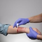
Most of the scholarly research on Centella has revealed utility in hyperproliferative skin conditions such as scarring and keloid formation. Asiaticoside, asiatic acid, and madecassic acid are among the triterpenoid wound-healing compounds in Centella and have been shown to help reduce excessive fibrosis in situations of scleroderma, extensive scar formation, and keloids.3 Centella has also been shown useful in promoting epithelial regeneration following burns.4 Proliferation of epithelial cells and angiogenesis are promoted by Centella. The expression of basic fibroblastic growth factor, an angiogenic factor, was also upregulated in tissues.5 Centella has been shown to promote the expression of genes involved in angiogenesis, growth factors, and extracellular hyaluronic acid-binding proteins, providing further explanation for the plant’s wound healing effects.6 Other investigations report that Centella promotes granular tissue, the strength of the granular tissue, and hydroxyproline content in both normal situations and steroid-suppressed healing response.7
Due to these angiogenesis promoting effects, Centella is also noted to improve microcirculation in the skin and might be included in formulas for poor wound healing, such as diabetics and patients with circulatory insufficiency.8,9 Improved venoarteriolar response, blood gases, capillary permeability, and general microcirculation have been reported in clinical trials.9
Although Centella is appropriate for promoting connective tissue formation, it is also appropriate for those with a tendency toward excessive connective tissue and keloid formation by normalizing appropriate collagen synthesis and reducing excessive fibrosis.10-12 The saponin asiaticoside induces type I collagen synthesis in human dermal fibroblast cells.10-12
Hypericum perforatum (St. John’s wort)
Hypericum is well known to promote photophobia with oral dosing and to have a photosensitizing effect when used topically, yet Hypericum paradoxically is shown to protect the skin from radiation damage. Hypericum flavonoids hypericin and hyperforin have been noted to limit the proliferation of T lymphocytes in the skin following radiation exposure, and yet hypericin is one of the most potent photosensitizers found in nature.13 A number or researchers have been investigating the photosensitizing ability of Hypericum as a therapy for mucosal and skin cancers. One recent study reported Hypericum to be a possible “photodynamic” therapy for nonmelanoma skin cancer.14 Hypericum is noted to promote collagen production by fibroblasts.15 The Hypericum flavanone astilbin is also reported to enhance hair growth via a mechanism likely involving transforming growth factor beta.16
Aloe Vera
Aloe vera has also been shown to support glycohydrolases, enzymes involved with the synthesis of glycosaminoglycans (GAGs) in the body, from which skin and bone are derived.17 The formation of both skin and bone involve the spontaneous organization of a complex, living, soluble, and electromagnetically active architecture first, which then becomes ossified in the case of bone, and “collagenized” in the case of skin. The dynamic organization matrix is sometimes referred to as the “ground substance” and is composed of GAGs which “granulate” with the synthesis of more solid collagen and elastin by fibroblasts. Both topical and internal use of Aloe vera promotes the formation of ground substance associated with measurable increases in hyaluronic acid and dermatan sulphate.17
Allantoin, an imidazole alkaloid found in Aloe (and Symphytum), acts as a cell-proliferating agent and has a GAG-like chemical makeup. Allantoin is produced endogenously by animals, is found in the milk of lactating animals, and appears to be involved with rapid growth. It is observed to increase in the serum of pregnant women and is decreased in cases of placental insufficiency.18
Hamamelis virginiana (Witch hazel)
Hamamelis is often overlooked by pigeonholing it as a hemorrhoid herb, but research has shown numerous wound healing and skin supportive effects from Hamamelis. Pain and swelling in wounds can often be allayed by Hamamelis soaks.19 Clinical investigations have shown Hamamelis to have a weak but noticeable ability to speed recovery of the skin following exposure to UV light.20,21 Hamamelis contains tannins, proanthocyanidins, and the polysaccharides arabans and arabinogalactans. Hamamelis proanthocyanidins have been shown to increase skin cell proliferation, which is an explanation for its long-standing folkloric reputation for relief of skin irritation.22 Hamamelis proanthocyanidins have also been shown to have activity against herpes simplex virus type I, thus it might be included in topical preparations for cold sores.23
Calendula (Pot Marigold)
Despite the fact that Calendula is perhaps the number one herb that herbalists think of for the skin, there are very few published studies regarding the physiologic effects of Calendula on the skin. One very recent study reported Calendula has an ability to reduce UV radiation-induced oxidative stress in the skin.24 Calendula has been reported to alleviate allergic inflammation of the hands and feet associated with chemotherapy side effects, and to reduce radiation-induced skin inflammation.25,26 Triterpenoid compounds in Calendula are credited with anti-inflammatory activity.27 Anti-edema activity has been reported from Calendula as evidence of preventing inflammation following exposure to irritants.28
To Be Continued
This discussion will continue in 2 months in the July issue of NDNR. Additional botanicals and some formula ideas will be covered.
 Jillian Stansbury, ND has practiced in SW Washington for nearly 20 years, specializing in women’s health, mental health and chronic disease. She holds undergraduate degrees in medical illustration and medical assisting, and graduated with honors in both programs. Dr. Stansbury also chairs the botanical medicine program at NCNM and teaches the core botanical curricula, a position she has held for over 18 years. In addition, Dr. Stansbury also writes and serves as a medical editor for numerous professional journals and lay publications, plus teaches natural products chemistry and herbal medicine around the country. At present she is working to set up a humanitarian service organization in Peru and studying South American ethnobotany. She is the mother of two adult children, and her hobbies include art, music, gardening, camping, international travel, and studying quantum and metaphysics.
References
- Gilliver SC, Ashcroft GS. Sex steroids and cutaneous wound healing: the contrasting influences of estrogens and androgens. Climacteric. 2007;10(4):276-288.
- Verdier-Sévrain S. Effect of estrogens on skin aging and the potential role of selective estrogen receptor modulators. Climacteric. 2007;10(4):289-297.
- Hong SS, Kim JH, Li H, Shim CK. Advanced formulation and pharmacological activity of hydrogel of the titrated extract of C. asiatica. Arch Pharm Res. 2005;28(4):502-508.
- Salas Campos L, Fernándes Mansilla M, Martínez de la Chica AM. Topical chemotherapy for the treatment of burns [in Spanish]. Rev Enferm. 2005;28(5):67-70.
- Cheng CL, Guo JS, Luk J, Koo MW. The healing effects of Centella extract and asiaticoside on acetic acid induced gastric ulcers in rats. Life Sci. 2004;74(18):2237-2249.
- Coldren CD, Hashim P, Ali JM, Oh SK, Sinskey AJ, Rha C. Gene expression changes in the human fibroblast induced by Centella asiatica triterpenoids. Planta Med. 2003;69(8):725-732.
- Shetty BS, Udupa SL, Udupa AL, Somayaji SN. Effect of Centella asiatica L (Umbelliferae) on normal and dexamethasone-suppressed wound healing in Wistar Albino rats. Int J Low Extrem Wounds. 2006;5(3):137-143.
- Wollina U, Abdel-Naser MB, Mani R. A review of the microcirculation in skin in patients with chronic venous insufficiency: the problem and the evidence available for therapeutic options. Int J Low Extrem Wounds. 2006;5(3):169-180.
- Cesarone MR, Incandela L, De Sanctis MT, et al. Evaluation of treatment of diabetic microangiopathy with total triterpenic fraction of Centella asiatica: a clinical prospective randomized trial with a microcirculatory model. Angiology. 2001;52(suppl 2):S49-S54.
- Lee J, Jung E, Kim Y, et al. Asiaticoside induces human collagen I synthesis through TGFbeta receptor I kinase (TbetaRI kinase)-independent Smad signaling. Planta Med. 2006;72(4):324-328.
- Lu L, Ying K, Wei S, et al. Asiaticoside induction for cell-cycle progression, proliferation and collagen synthesis in human dermal fibroblasts. Int J Dermatol. 2004;43(11):801-807.
- Lu L, Ying K, Wei S, Liu Y, Lin H, Mao Y. Dermal fibroblast-associated gene induction by asiaticoside shown in vitro by DNA microarray analysis. Br J Dermatol. 2004;151(3):571-578.
- Schempp CM, Winghofer B, Lüdtke R, Simon-Haarhaus B, Schöpf E, Simon JC. Topical application of St John’s wort (Hypericum perforatum L.) and of its metabolite hyperforin inhibits the allostimulatory capacity of epidermal cells. Br J Dermatol. 2000;142(5):979-984.
- Kacerovská D, Pizinger K, Majer F, Smíd F. Photodynamic therapy of nonmelanoma skin cancer with topical hypericum perforatum extract–a pilot study. Photochem Photobiol. 2008;84(3):779-785.
- Oztürk N, Korkmaz S, Oztürk Y. Wound-healing activity of St. John’s Wort (Hypericum perforatum L.) on chicken embryonic fibroblasts. J Ethnopharmacol. 2007;111(1):33-39.
- Sasajima M, Moriwaki S, Hotta M, Kitahara T, Takema Y. trans-3,4′-Dimethyl-3-hydroxyflavanone, a hair growth enhancing active component, decreases active transforming growth factor beta2 (TGF-beta2) through control of urokinase-type plasminogen activator (uPA) on the surface of keratinocytes. Biol Pharm Bull. 2008;31(3):449-453.
- Chithra P, Sajithlal GB, Chandrakasan G. Influence of Aloe vera on the glycosaminoglycans in the matrix of healing dermal wounds in rats. J Ethnopharmacol. 1998;59(3):179-186.
- Shestopalov AV, Shkurat TP, Mikashinovich ZI, et al. Biological functions of allantoin [in Russian]. Izv Akad Nauk Ser Biol. 2006;5:541-545.
- Wolff HH, Kieser M. Hamamelis in children with skin disorders and skin injuries: results of an observational study. Eur J Pediatr. 2007;166(9):943-948.
- Hughes-Formella BJ, Filbry A, Gassmueller J, Rippke F. Anti-inflammatory efficacy of topical preparations with 10% hamamelis distillate in a UV erythema test. Skin Pharmacol Appl Skin Physiol. 2002;15(2):125-132.
- Korting HC, Schäfer-Korting M, Hart H, Laux P, Schmid M. Anti-inflammatory activity of hamamelis distillate applied topically to the skin. Influence of vehicle and dose. Eur J Clin Pharmacol. 1993;44(4):315-318.
- Deters A, Dauer A, Schnetz E, Fartasch M, Hensel A. High molecular compounds (polysaccharides and proanthocyanidins) from Hamamelis virginiana bark: influence on human skin keratinocyte proliferation and differentiation and influence on irritated skin. Phytochemistry. 2001;58(6):949-958.
- Erdelmeier CA, Cinatl J Jr, Rabenau H, Doerr HW, Biber A, Koch E. Antiviral and antiphlogistic activities of Hamamelis virginiana bark. Planta Med. 1996;62(3):241-245.
- Fonseca YM, Catini CD, Vicentini FT, Nomizo A, Gerlach RF, Fonseca MJ. Protective effect of Calendula officinalis extract against UVB-induced oxidative stress in skin: evaluation of reduced glutathione levels and matrix metalloproteinase secretion. J Ethnopharmacol. 2010;127(3):596-601.
- Kern E, Schmidinger M, Locker GJ, Kopp B. Management of capecitabine-induced hand-foot syndrome by local phytotherapy [in German]. Wien Med Wochenschr. 2007;157(13-14):337-342.
- Chargari C, Fromantin I, Kirova YM. Importance of local skin treatments during radiotherapy for prevention and treatment of radio-induced epithelitis [in French]. Cancer Radiother. 2009;13(4):259-266.
- Neukirch H, D’Ambrosio M, Sosa S, Altinier G, Della Loggia R, Guerriero A. Improved anti-inflammatory activity of three new terpenoids derived, by systematic chemical modifications, from the abundant triterpenes of the flowery plant Calendula officinalis. Chem Biodivers. 2005;2(5):657-671.
- Zitterl-Eglseer K, Sosa S, Jurenitsch J, et al. Anti-oedematous activities of the main triterpendiol esters of marigold (Calendula officinalis L.). J Ethnopharmacol. 1997;57(2):139-144.


