Exploring the Role of Tumor Microenvironment in Prostate Cancer and the Potential in Natural Therapies
By Fraser Smith
This article examines the role of the extracellular matrix in prostate cancer progression and highlights natural therapies that target the tumor microenvironment.
Understanding Prostate Cancer
Prostate cancer is a highly prevalent condition. According to the Centers for Disease Control, out of 100 men, 13 will develop prostate cancer during their lifetime, and two to three will die from it.1 It can emerge in earlier years and spread quickly (often to the bone – the spine, sacrum, and even appendicular skeleton). Interestingly, autopsies show that many men in their 80s harbor microtumors that never progressed to cause harm.
Risk factors for prostate cancer include genetic predisposition, familial history, age, and race, with, African-Americans experiencing a higher prevalence. Exposure to endocrine disruptors, in which our society is awash, can have proliferative effects on prostate cancer cells by activating the androgen receptors.2Diagnosis typically involves prostate biopsies and Gleason scoring, which grades the cancer on a scale from one to 10 based on tissue differentiation and aggressiveness.3 Imaging, such as MRI is often done as well.
Conventional Treatments for Prostate Cancer
There are medical treatments for prostate cancer including surgery, various forms of radiation delivery, and endocrine therapy.4 Mostly, physical (surgical, cryotherapy, thermal therapy) or energetic ablation and the suppression of pro-proliferative androgens have been the successful treatments. This sets prostate cancer apart in the sense that cytotoxic approaches have not been used except in metastases when drugs such as Docetaxel might be used.
Emerging treatments include engineered immune cells and antibody therapies that direct the immune system’s attention toward prostate cancer.
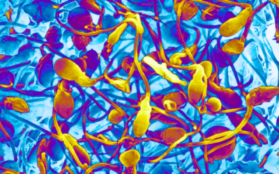
The Role of the Extracellular Matrix (ECM) in Cancer Progression
A cancer cell’s ability to dominate its immediate environment, gain increasing access to nutrients, and spread locally relies on key functions of the tumor microenvironment. This complex milieu is largely shaped by the extracellular matrix (ECM) and its interactions with the cancer cells. The ECM serves as a critical foundation for cellular viability and health, And our understanding of its architecture and its processes has grown rapidly. As naturopathic physicians who attempt to adjust the terrain in addition to confronting specific pathologies and problems, considering the ECM is of importance.
The ECM used to be commonly referred to as the “ground substance” and this term is still used concerning the hyaluronic acid components of the ECM that create a gel structure. This was as far as many histology courses got in appreciating this vastly important regulatory structure. The ECM is rich with proteins, and proteoglycans.5, 6 Proteins including fibrillar and non-fibrillar collagen (composed of three polypeptide α chains) and elastin (composed of elastin and fibrillin). Collagen has fundamental structural importance, and elastin gives the matrix tensile strength, allowing it to withstand stress and compressional forces from different directions.
Proteoglycans are created by binding a protein to glycosaminoglycans (GAG), which are made from repeating disaccharide units. GAGs can be hyaluronic acid (the only non-sulfated GAG), keratan sulfate; chondroitin/dermatan sulfate; and heparan sulfate, including heparin. All except hyaluronic acid are sulfated. These have a negative charge, which attracts water and cations and provides bulk to the ECM.
Proteoglycans have some similarities to glycoproteins but are essentially glycosaminoglycans (such as chondroitin sulfate) with a protein side chain. These molecules have a structural role but are vastly important in controlling cell signaling and deploying growth factors. Examples are perlecan, aggrecan, and syndecan. Small leucine-rich proteoglycans are associated with collagen and play a major role in the organization of collagenous structures (they are found in great numbers in the cornea and all tissues).
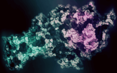
The protein integrin is found in all cells and acts as a connector between the proteins of the ECM and the cell. Integrin is a transmembrane protein, and on the ECM side, another protein known as fibronectin connects integrin to ECM proteins. On the intracellular side, integrin is connected to the microtubule structure of the cell. Those microtubules connect with the cell nucleus. Mechanical signals from the ECM are translated, via integrin, to the cytoskeleton and ultimately the nucleus. This has a well-demonstrated effect on gene transcription.
As we look at each of these categories, we find more specialized types of ECM molecules and it becomes clear that the ECM is a central actor in biological function. It is not surprising then, that the battle of a cancer cell to overcome host defenses and infiltrate its environment is fought within, and over control of, the ECM.
ECM Dysregulation in Prostate Cancer
Neoplastic cells are very active in their micro-environment, which is the extracellular matrix, basement membranes, and the nearby cells of the matrix, including fibroblasts and macrophages.7 One of the early changes is that under the influence of the neoplastic cells, certain fibroblasts start to lay down excessive amounts of collagen. This deposition of collagen is just the first step, but an important one. The term “tumor-associated fibroblasts” refers to those cells that are now working for the neoplastic cells.8
Once this deposition is underway, the collagen meshwork, which has become unusually dense, is modified. The particular modification is to crosslink the collagen. Some crosslinking naturally happens in collagen, which is important for stability. Tissues that must withstand more stress, might display more cross-linking. Under the influence of neoplastic cells, the local collagen becomes excessively crosslinked. This provides several benefits to the cancer cells. One is that it can provide cover from circulating natural killer (NK) cells. It also leads to a rigid proximal environment to the cancer cell. This has direct effects on the cancer cell nucleus through the mechanotransduction effects of this collagen border. Especially, it has a proliferative effect.
Indeed, the stiffening of prostate tissue in prostate cancer is associated with prostate cancer progression. Prostate cancers that are not responsive to endocrine-suppressing therapies can become treatment-resistant, and metastasize. The stiffer the tumor, the more likely it is going to progress and spread.9, 10, 11
Cancer cells don’t merely divide, they invade. In some tumors, especially around a basement membrane, the tumor cells form “invadopodia.” These are extensions of the cell that are studded with matrix metalloproteinases (MMP). These enzymes dissolve matrix proteins and are part of the normal maintenance of the ECM. However, under the direction of cancer cells, fibroblasts can be induced to produce excessive amounts of MMP. The invadopodia can penetrate the microenvironment, and break through normal stromal and basal laminal boundaries.
Cancer cells do not stay in one place. They attempt to move through the extracellular matrix. They use the integrin and fibronectin molecules to migrate through the ECM, even as they are “tunneling” through aspects of it. This massive destruction doesn’t only create pathways for new cancer cells, it releases stored growth hormones from vesicles with the ECM. These are normally released as needed, but the wholesale destruction of the ECM leads to their sudden release. This further accelerates tumor growth. Some of these growth factors are angiogenic, thus encouraging the sprouting of new blood vessels into the area. This brings more nutrients, and, of course, more opportunities to spread cancer cells.
Natural therapies Targeting the ECM
Many natural therapies can work on, and perhaps through the ECM. Manual therapy, electroacupuncture, and hydrotherapy all have demonstrated effects on the ECM. Concerning tumor strategies that dominate and exploit the microenvironment, certain plant extracts have been shown to work against these biochemical moves made by neoplastic cells.
Milk Thistle (Silybum marinaum)
Silibinin is a flavonolignan that is derived from Silybum marinaum, or Milk Thistle. Widely used for its antioxidant and hepatoprotective functions, it has actions in the ECM. According to Liang et al7, silibinin interferes with the interaction between prostate cancer cells and fibronectin, inhibiting their motility, invasiveness, and survival. Deep et al, in a 2014 paper in Mutation Research, have demonstrated that silibinin modulates fibronectin-induced expression of integrins (α5, αV, β1, and β3), actin remodeling (e.g., local adhesion kinase), apoptosis, EMT, and signaling molecules related to cell survival.12
Turmeric (Curcuma longa)
The polyphenol curcumin is derived from Curcuman longa or turmeric. Its use as a dietary supplement is extensive. It can suppress the cancer (or tumor) associated fibroblasts that create the dense collagen that prostate cancer cells thrive in. Zeng et al found that curcumin effectively increases the intracellular Reactive oxygen species levels of prostate-Cancer Associated Fibroblasts and promotes their apoptosis and cell cycle arrest in G2-M phase.
Du et al, using a cell culture model, found that cancer-associated fibroblasts (CAF) can induce prostate cancer cells to differentiate further, becoming more mesenchyme than epithelial cells. These CAFs can also help prostate cancer cells become more invasive. They do this via a monoamine oxidase A (MAOA)/mammalian target of rapamycin (mTOR)/hypoxia-inducible factor-1α (HIF-1α) signaling pathway, which exploits reactive oxygen species (ROS) to drive a migratory and aggressive phenotype of prostate carcinoma cells. Du et al also found that CAF was able to increase CXC chemokine receptor 4 (CXCR4) and interleukin-6 (IL-6) receptor expression in prostate cancer cells. Once again, curcumin seems to fight against these cancer strategies. It stopped the invasion into the immediate environment, caused by CAF, and also stopped the transition from epithelium to mesenchyme. It inhibited MAOA/mTOR/HIF-1α signaling which lowered inflammatory cytokine IL-6. This all together slowed down the prostate cancer cells in this model.
Green Tea (Camellia sinesis)
Epicatechin-3-gallate (ECG), EGCG, and catechins are found in Camellia sinensis or green tea. Lang et al note that ECG suppresses cancer invasiveness. Catechin reduces excessive matrix metalloproteinase activity, the enzymes used to reshape or destroy parts of the ECM during cancer spread. EGCG inhibits prostate CAFs differentiation, which means less excessive, crosslinked, and tumor-stimulating collagen in the ECM.
Gale of the Wind (Phallanthus amarus)
Phallanthus amarus or Gale of the Wind, which has uses in many traditional medicine systems, has been found to reduce prostate cancer invasiveness.15 In a matrix model, Tang et al found that Phyllanthus extracts significantly inhibited cell adhesion, migration, invasion, and transendothelial migration activities of cancer.16 The Phyallanthus extracts also had an anti-angiogenic effect (important to reduce new blood supply to the cancer cells) and reduced excessive matrix metalloproteinase activity caused by prostate cancer cells.
Conclusion
There are many aspects to the treatment of prostate cancer and optimum support for the patients who are fighting it. The use of naturopathic therapeutics goes beyond the tumor microenvironment. However, the events that take place in the extracellular matrix near the cancer site are so integral to prostate cancer spread, that the promising effects of naturopathic therapies, particularly plant extracts, deserve attention. They may well have the ability to restore the unbalanced microenvironment to a properly regulated and balanced one, which tips the scales in our favor.
Case Study
A 74-year-old male presented to his urologist with findings of unusual stiffness during a digital rectal examination (DRE) A prostate-specific antigen (PSA) test was subsequently ordered, revealing a PSA level of 15ng/mL. Further diagnostic workup, including an MRI of the prostate and pelvis and a biopsy, confirmed a diagnosis of prostate adenocarcinoma. The cancer was graded as a Gleason score of 7 (3+4) and classified as a Grade Group II (Stage II C), with no evidence of metastasis beyond the prostate.
Given the localized nature of the cancer and its intermediate risk, surgery was not recommended at this time. Instead, the patient was advised to engage in surveillance with regular monitoring.
During a comprehensive intake by his naturopathic physician, it was noted that the patient’s diet, while free from most “fast food,” was deficient in plant-based foods, particularly fruits and vegetables. Dietary recommendations were made to address this gap, focusing on sustainable changes to shopping habits and meal preparation to increase the intake of nutrient-dense plant foods. To further support the patient’s condition, the naturopathic physician prescribed a supplement containing curcumin and quercetin nanoparticles.17 He hopes that these nano packages of curcuminoids and quercetin are more bioavailable, not only via absorption from the gut but transit to and through the extracellular matrix in the prostate tissue. The ND adds to this an isoflavone supplement, made from Trifolium pratense (red clover).18 Meanwhile, they advise the patient to avoid food stored in plastic as much as possible and never cook or store food in plastic to avoid xenoestrogens, which could have a dysregulating effect on the prostate tissue. They consider future therapies and review the importance of keeping up with all monitoring actions suggested by the urologist. The ND performs separate DRE and PSA tests as part of ongoing care but expects the patient to continue his work with the urologist.

Fraser Smith, MATD, ND is a Professor of Clinical Sciences at National University of Health Sciences in Lombard, Illinois, where he has led the ND program since its launch in 2006. He is the author of textbooks and nutrition books for the public, including Naturopathic Medicine: A Comprehensive Guide (Springer, 2022).
References
1. Prostate Cancer Risk Factors. Centers for Disease Control. https://www.cdc.gov/prostate-cancer/risk-factors/index.html Accessed 10/20/2024.
2. Corti M, Lorenzetti S, Ubaldi A, Zilli R, Marcoccia D. Endocrine Disruptors and Prostate Cancer. Int J Mol Sci. 2022;23(3):1216. Published 2022 Jan 21. doi:10.3390/ijms23031216
3. Sehn JK. Prostate Cancer Pathology: Recent Updates and Controversies. Mo Med. 2018;115(2):151-155.
4. Sekhoacha M, Riet K, Motloung P, Gumenku L, Adegoke A, Mashele S. Prostate Cancer Review: Genetics, Diagnosis, Treatment Options, and Alternative Approaches. Molecules. 2022;27(17):5730. Published 2022 Sep 5. doi:10.3390/molecules27175730
5. Karamanos NK, Theocharis AD, Piperigkou Z, et al. A guide to the composition and functions of the extracellular matrix. FEBS J. 2021;288(24):6850-6912. doi:10.1111/febs.15776
6. Yue B. Biology of the extracellular matrix: an overview. J Glaucoma. 2014 Oct-Nov;23(8 Suppl 1):S20-3. doi: 10.1097/IJG.0000000000000108. PMID: 25275899; PMCID: PMC4185430.
7. Liang D, Liu L, Zhao Y, et al. Targeting extracellular matrix through phytochemicals: a promising approach of multi-step actions on the treatment and prevention of cancer. Front Pharmacol. 2023;14:1186712. Published 2023 Jul 25. doi:10.3389/fphar.2023.1186712
8. Biffi G, Tuveson DA. Diversity and Biology of Cancer-Associated Fibroblasts. Physiol Rev. 2021;101(1):147-176. doi:10.1152/physrev.00048.2019
9. Luthold C, Hallal T, Labbé DP, Bordeleau F. The Extracellular Matrix Stiffening: A Trigger of Prostate Cancer Progression and Castration Resistance?. Cancers (Basel). 2022;14(12):2887. Published 2022 Jun 11. doi:10.3390/cancers14122887
10. Stewart DA, Cooper CR, Sikes RA. Changes in extracellular matrix (ECM) and ECM-associated proteins in the metastatic progression of prostate cancer. Reprod Biol Endocrinol. 2004;2:2. Published 2004 Jan 7. doi:10.1186/1477-7827-2-2
11. Kang J, La Manna F, Bonollo F, et al. Tumor microenvironment mechanisms and bone metastatic disease progression of prostate cancer. Cancer Lett. 2022;530:156-169. doi:10.1016/j.canlet.2022.01.015
12. Deep G, Kumar R, Jain AK, Agarwal C, Agarwal R. Silibinin inhibits fibronectin induced motility, invasiveness and survival in human prostate carcinoma PC3 cells via targeting integrin signaling. Mutat Res. 2014;768:35-46. doi:10.1016/j.mrfmmm.2014.05.002
13. Zeng Y, Du Q, Zhang Z, et al. Curcumin promotes cancer-associated fibroblasts apoptosis via ROS-mediated endoplasmic reticulum stress. Arch Biochem Biophys. 2020;694:108613. doi:10.1016/j.abb.2020.108613
14. Du Y, Long Q, Zhang L, et al. Curcumin inhibits cancer-associated fibroblast-driven prostate cancer invasion through MAOA/mTOR/HIF-1α signaling. Int J Oncol. 2015;47(6):2064-2072. doi:10.3892/ijo.2015.3202
15. Salehi B, Fokou PVT, Yamthe LRT, Tali BT, Adetunji CO, Rahavian A, Mudau FN, Martorell M, Setzer WN, Rodrigues CF, Martins N, Cho WC, Sharifi-Rad J. Phytochemicals in Prostate Cancer: From Bioactive Molecules to Upcoming Therapeutic Agents. Nutrients. 2019 Jun 29;11(7):1483. doi: 10.3390/nu11071483. PMID: 31261861; PMCID: PMC6683070.
16. Tang YQ, Jaganath IB, Manikam R, Sekaran SD. Phyllanthus spp. Exerts Anti-Angiogenic and Anti-Metastatic Effects Through Inhibition on Matrix Metalloproteinase Enzymes. Nutr Cancer. 2015;67(5):783-795. doi:10.1080/01635581.2015.1040518
Case specific references
17. Turkekul K, Erdogan S. Potent Suppression of Prostate Cancer Cell Growth and Eradication of Cancer Stem Cells by CD44-targeted Nanoliposome-quercetin Nanoparticles. J Cancer Prev. 2023 Dec 30;28(4):160-174. doi: 10.15430/JCP.2023.28.4.160. PMID: 38205358; PMCID: PMC10774486.
18. Zhang HY, Cui J, Zhang Y, Wang ZL, Chong T, Wang ZM. Isoflavones and Prostate Cancer: A Review of Some Critical Issues. Chin Med J (Engl). 2016;129(3):341-347. doi:10.4103/0366-6999.174488