

A chronic costal-margin pain case highlights how neurodynamic testing and reproduction of concordant symptoms can reveal a thoracic neural source missed by visceral workups and static imaging—and guide targeted orthobiologic treatment.
Thoracic radicular pain can present atypically, confined to part of a dermatome along the chest or abdominal wall rather than involving the full dermatomal distribution, which can lead to unnecessary workups and delays in treatment.
Case presentation
A 43-year-old man developed right anterolateral costal margin “pinching” that progressed to a “clawing” pain, with mid-thoracic back pain emerging months later. Symptoms were provoked within minutes of driving and by loaded flexion, and refractory to extensive conservative care. Visceral and chest-wall investigations were unremarkable.
Intervention
After comprehensive physical examination including clinical neurodynamic testing and point-of-care ultrasound, a single image-guided platelet-based session targeted the right T10 nerve root via the T10-T11 transforaminal epidural space, right T8-T9 through T10-T11 facet joints, the right T9-T10 costotransverse joint, and the lateral cutaneous branch of the 10th intercostal nerve and associated fascia at the costal margin.
Outcomes
Pain was rated on an 11-point (0-10) numeric rating scale, where higher is worse. Six months after treatment, usual pain improved from 6 to 1, and current pain from 4 to 0. Desired functionality, rated 0 to 100 where higher is better, rose from 85 at baseline to 100 at 1 and 3 months, and was 90 at 6 months. The Oswestry Disability Index was low at baseline and unchanged at 6 months, 6 in both cases. Additionally, the patient reported being extremely satisfied at 6 months.
Conclusion
When visceral workup was unrevealing, repeated reproduction of concordant symptoms through examination and neurodynamic assessment informed a working thoracic localization that static imaging did not identify. In this patient, sustained improvement was observed after a targeted multimodal platelet-based intervention.
Keywords: thoracic radiculopathy; intercostal neuralgia; costal margin pain; platelet-rich plasma; neurodynamic testing
Introduction
Thoracic radicular syndromes receive far less attention than their cervical and lumbar counterparts, and the interventional literature addressing them is sparse.1 Diagnosis is complicated by the region’s overlapping somatic, radicular, visceral, and referred pain sources. Additionally, abdominal-wall pain is frequently mistaken for visceral disease and prompts extensive negative gastrointestinal evaluations.2,3 Imaging can add to the complexity, since degenerative thoracic findings may or may not be symptomatic, and therefore must be correlated with the clinical presentation before they are accepted as the pain generator. Platelet-based injections for radicular pain are an emerging area of study, however certainty remains limited.4
This report describes an unusual presentation of chronic thoracic radicular pain dominated by right anterolateral costal margin symptoms (simply referred to as costal margin symptoms in this report). It illustrates how neurodynamic testing, together with recreation of the patient’s familiar (concordant) pain on palpation, localized the pain source and directed treatment when two years of visceral and MRI-based thoracic spine investigation could not.
Case Presentation
Patient Information
A 43-year-old physically active male whose occupation requires prolonged driving presented with chronic right-sided costal margin pain two years in duration. Prior to onset, pertinent medical history was unremarkable for any spinal, visceral, or abdominal pathology. He took no relevant medications.
Presenting Concern
Symptoms began in June 2023 as a right lower-rib-region “pinching,” evolving into a “clawing” sensation under the costal margin. Six to eight months following the onset of his symptoms, he developed right mid-thoracic back pain. The anterolateral costal margin pain, rather than the thoracic pain, was the most disabling feature and was the primary driver for seeking diagnostic workup and treatment at our clinic.
The patient’s symptoms were provoked and exacerbated with specific positions, supporting a musculoskeletal etiology. Specifically, within 4-5 minutes of driving his costal margin clawing would appear, which would flare to 8-9/10 with extended driving, but would significantly decrease upon standing. Additionally, the patient reported that loaded flexion, such as resistance cycling, planks, and dumbbell rows, was provocative, while standing, extension maneuvers, and direct pressure over the anterolateral costal margin provided relief.
Clinical History and Context
Of importance, the onset coincided with a period of acute stress following his father’s death from a gastrointestinal illness, which had prompted particular concern for a visceral etiology. This led to two years of extensive visceral and chest-wall evaluations, but these were unrevealing. The patient exhausted conservative care, including physical therapy with four providers over approximately one year, two courses of chiropractic care, massage, dry needling, topical diclofenac, and ibuprofen. All conservative approaches provided temporary or no relief. He declined a prescription for gabapentin due to side effect concerns, and a 3-day SSRI trial was stopped due to intolerance.
Clinical Findings
An initial telehealth neurodynamic screen in October 2025 was conducted, which found the following: Self-performed seated Bechterew’s test on the right was negative, but adding cervical flexion recreated concordant symptoms in the costal margin. In that position, contralateral knee extension and ankle dorsiflexion reduced these concordant symptoms. Additionally, while in the dumbbell row position, he reported concordant symptoms in both the anterolateral costal margin and right lower thoracic spine; in this position, cervical flexion increased the right thoracic spine pain, and cervical extension reduced pain in both the costal margin and right thoracic spine.
A thorough physical examination was performed in office in November 2025, which elicited concordant tenderness over the right T8-T9 through T10-T11 facet joints and the right T9-T10 costotransverse joint, while concordant pain on digital sonopalpation of the intertransverse space between T10 and T11 supported a working T10-level root localization. Key physical examination findings are presented in Table 2.
Diagnostic Assessment
The diagnostic workup and subsequent care are summarized chronologically in Table 1. CT enterography in July 2023 showed no inflammatory or visceral pathology. Contrast abdominal MRI with magnetic resonance cholangiopancreatography in December 2024 identified no explanatory lesion, and targeted right chest-wall ultrasound in January 2025 was unremarkable. The patient had four in-office abdominal-wall ultrasounds. Outside reports for two of these (December 2024 and May 2025) documented grade 2 strains with partial tears of the right rectus abdominis beneath the 12th rib and of the medial right external oblique, unchanged at follow-up, with normal internal oblique and transversus abdominis and intact connecting fascia; the others were unremarkable per the patient’s report. On the author’s review of the reports and images, these tears were not at the site of the fascial-plane finding described below.
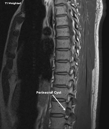
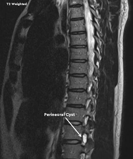
Thoracic MRI in June 2025 showed a mild convex-right mid-thoracic curvature, minuscule disc protrusions at T1-2, T3-4, T5-6 (with annular fissure), and T10-11, all without canal compromise or cord signal abnormality, and a tiny right T12-L1 foraminal perineural cyst (Figures 1 and 2).
Point-of-care ultrasound at the November 2025 visit showed mild effusion and cortical irregularity at the right T8-T9 facet joint, a sonographically unremarkable T9-T10 costotransverse joint that was tender on sonopalpation with concordant pain, and a discrete anechoic layer between the external and internal oblique just inferior to the 10th costal margin (Figure 3), over which sonopalpation reproduced the costal margin symptoms.
Standardized patient-reported outcome surveys were administered through a third-party registry, which used the 0-10 numeric rating scale for pain (higher is worse), a 0-100 desired-functionality scale (higher is better), and the Oswestry Disability Index overall score (higher indicates greater disability). Survey results are found in Table 5.
“Persistent anterolateral costal margin pain may reflect a thoracic neuropathic process especially when abdominal and chest-wall investigations are unrevealing.”
Clinical Assessment
The working diagnosis was T10 radicular pain, with concomitant right thoracic facet and costotransverse arthralgia and possible neuralgia involving the lateral cutaneous branch of the 10th intercostal nerve. Nerve root or dural involvement was supported by slump testing reproducing concordant thoracic and costal margin pain, both of which were altered with change in cervical position, and palpation between the right T10 and T11 transverse processes recreating the costal margin pain. Concordant tenderness at the T8-T9 through T10-T11 facet joints and the T9-T10 costotransverse joint suggested these were contributing to or resulting from the nerve irritation, and sonopalpation over the anechoic layer recreated the costal margin symptoms, raising suspicion for a possible double crush syndrome.5 Given its size and irregular shape, the anechoic layer could also represent a fascial tear.
Therapeutic Intervention
The previously identified pain generators were treated with autologous platelet-based products under image guidance in November 2025; the full details can be found in Table 3. Briefly, a T10-T11 infraneural transforaminal epidural was performed under fluoroscopy with contrast confirmation (Figures 4 and 5), where 2 mL of a planned 4 mL mixture of platelet-rich plasma (PRP) and platelet lysate (PL), prepared as 1 mL PRP with 3 mL PL, was injected. The final volume was reduced due to intense pressure reported by the patient. The T8-T9 through T10-T11 facet joints and the T9-T10 costotransverse joint were then injected under ultrasound and fluoroscopic guidance with contrast confirmation, each receiving PRP intra-articular and periarticular, followed by a platelet-poor plasma solution (PPPS, a mixture of 13 mL platelet-poor plasma, 5% dextrose, and 0.25% ropivacaine) into the overlying multifidus and the muscle overlying the costotransverse joint. Lastly, ultrasound-guided fascial-plane hydrodissection inferior to the right 10th costal margin delivered 5 mL platelet lysate solution (PLS, a mixture of 8 mL platelet lysate, 5% dextrose, and 0.1% ropivacaine) between the internal and external oblique and 5 mL PPPS superficial to the external oblique.
The autologous preparations were made in-house from 240 mL whole blood containing 20% acid citrate dextrose solution A (ACD-A) by double-spin centrifugation, yielding 18 mL PRP with an 11.7-fold platelet enrichment and 87.5% recovery (Table 4). Total leukocytes were 1.3-fold baseline, comprising a 3.4-fold lymphocyte increase and an 88% granulocyte reduction, so by total count the preparation was leukocyte-enriched but granulocyte-poor and lymphocyte-rich. Platelet lysate was prepared from 11 mL of the PRP, approximately 19.6 billion platelets, by ultrasonic bath processing followed by serial 0.45/0.22 micron filtration to remove platelet debris, with 10.5 mL of lysate recovered after filtration.
Follow-up and Outcomes
The patient was followed by telephone at 3 days, 2 weeks, and 1 month, by telehealth visits at 3 months and approximately 8.5 months, and by standardized surveys at baseline, 1, 3, and 6 months. An early transient increase in right-upper-quadrant symptoms at 3 days settled without intervention, and at 2 weeks he described feeling “the best he had felt in a long time.” At 1 month, thoracic pain was subjectively 100% improved and costal margin symptoms approximately 75% improved. At 3 months, thoracic pain remained about 95% improved, he had completed a multi-day golf trip without thoracic symptoms and strength trained without restriction, and costal margin symptoms were about 70% improved. Compared to baseline, surveys at 6 months demonstrated improvements in usual pain (6/10 to 1/10), current pain (4/10 to 0/10), worst pain (9/10 to 4/10), and desired functionality (85/100 to 90/100), while the ODI remained stable at low disability (6 at both time points). Additionally, the patient reported he was “extremely satisfied” with his improvement. At his most recent telehealth follow-up (approximately 8.5 months post injection), thoracic pain was maintained at about 97% improved, while the costal margin symptoms fluctuated, with a window of several days of complete resolution and then a partial return described as annoying but not debilitating.
Safety and Tolerability
There were no immediate procedural complications. Beyond the transient 3-day flare, the only possible adverse event was a localized pruritic, hive-like eruption over the treatment sites 2 weeks post procedure, without fever, swelling, or signs of infection. This was managed with oral antihistamines and observation, and fully resolved shortly after onset. Causality remained uncertain given the delayed timing.
Timeline
Table 1. Chronological timeline of key events.
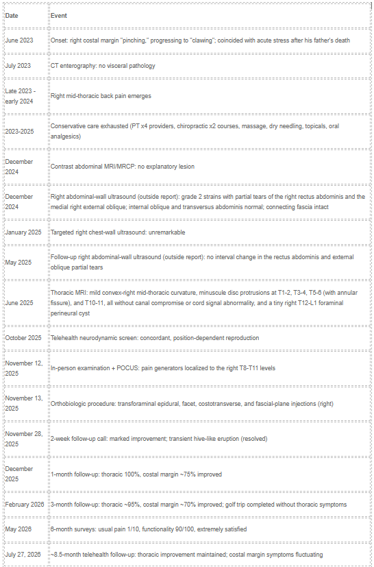
Abbreviations: CT, computed tomography; MRCP, magnetic resonance cholangiopancreatography; MRI, magnetic resonance imaging; POCUS, point-of-care ultrasound; PT, physical therapy.
Discussion
Radicular pain is uncommon in the thoracic spine relative to the cervical and lumbar spine, and clinicians have correspondingly less pattern exposure.1 The dominant complaint was pain at the anterolateral costal margin and right upper quadrant, therefore concern for gastrointestinal disease drove two years of negative visceral evaluation, which is typical of how thoracic radicular and abdominal-wall pain syndromes evade diagnosis.2,3
The diagnosis for this case rested on a comprehensive physical examination and reproduction of the patient’s concordant symptoms, in particular with clinical neurodynamic testing, rather than on static imaging. The prior MRI demonstrated a T12-L1 perineural cyst, which can cause radicular pain;6 however, this was deemed incidental and asymptomatic, as the history and physical examination demonstrated the pain to be originating from a more cephalad thoracic level. The MRI showed only a tiny posterior-left T10-T11 protrusion without central or foraminal stenosis, so no compressive lesion explained right T10 radicular pain, which can occur without demonstrable root compression.7
Neurodynamic testing was crucial in this patient’s workup, and the tissue movement behind it has been measured directly. Cadaveric dissection shows that the cord, dura, and nerve roots move within the spinal canal with flexion and extension of the head and neck, and that the stretch produced by cervical flexion involves the thoracic as well as the cervical segments of the cord.8 In vivo MRI in asymptomatic subjects shows that a straight leg raise displaces the conus caudally by about 3.5 mm, whereas a sham maneuver that preserves hip and pelvic motion while reducing neural tension produces no significant mean displacement, consistent with tensile-force transmission through the lumbosacral roots and dura.9 In the same model, bilateral straight leg raise produced nearly twice the caudal conus displacement of unilateral loading (4.58 versus 2.33 mm).10 Separately, in 88 asymptomatic adults, cervical position altered knee-flexion range during slump testing, while ankle dorsiflexion altered hip-flexion range during the straight leg raise.11 These findings provide a mechanical rationale for the case-specific observations that cervical position altered the distribution and intensity of symptoms during neurodynamic testing; those observations supported the diagnosis of nerve involvement.
As with many cases of chronic pain, the physical examination suggested more than one pain-generating tissue, with proximal articular and root-level findings at T8-T11 and a distal fascial-plane finding at the costal margin with concordant sonopalpation. A double-crush mechanism is one model that reconciles proximal and distal irritability of the same neural pathway,5 but true two-site pathology from a single primary generator with secondary sensitization could not be distinguished in this case.
Evidence for epidural platelet-rich plasma and platelet derivatives is emerging, mostly in the lumbar spine, with limited certainty,4,12,13 and facet-joint PRP data are limited with mixed results. Even so, the available studies suggest that platelet-based epidural injections can be effective: a 2025 meta-analysis of randomized trials found epidural PRP comparable to epidural steroid for lumbar radiculopathy,14 and an uncontrolled registry of 470 patients given epidural platelet lysate for lumbar radicular pain reported improvement through 24 months.15
Limitations. This is a single uncontrolled case, therefore natural history, regression to the mean, and placebo or belief effects cannot be excluded as the reason for improvement. Additionally, simultaneous treatment of multiple structures prevents attribution to any single tissue. Growth factor analysis was not performed on the lysate, so it is characterized only by the platelet dose prior to ultrasonication. All follow-ups were remote via telephone or video conference software, therefore a follow-up physical examination could not be performed to determine if the expected neurodynamic tests improved in conjunction with symptom improvement.
Conclusion
Persistent anterolateral costal margin pain may reflect a thoracic neuropathic process especially when abdominal and chest-wall investigations are unrevealing. In this case, repeated reproduction and modulation of concordant symptoms during neurodynamic testing and thoracic palpation was crucial in obtaining an accurate diagnosis, rather than the standard approach of simply obtaining an MRI. A targeted multi-structure platelet-based intervention was followed by substantial, durable improvement in pain for more than 8 months, with return to golf and strength training without restriction and high patient satisfaction.
Tables and Figures
Table 1. Chronological timeline of key events.
Table 2. Physical examination findings from the telehealth neurodynamic screen and the in-person examination.
Table 3. Treatment characteristics of the orthobiologic procedure, including target, guidance, and injectate.
Table 4. Biologic sample analysis of the whole blood and the platelet-rich plasma preparation.
Table 5. Standardized patient registry surveys from baseline through 6 months.
Figure 1. Sagittal T2-weighted thoracic MRI, June 2025, showing the right T12-L1 foraminal perineural cyst (arrow).
Figure 2. Sagittal T1-weighted thoracic MRI, June 2025, showing the same T12-L1 perineural cyst (arrow).
Figure 3. Point-of-care ultrasound of the right anterolateral costal margin at the 10th rib, showing the discrete anechoic layer between the external and internal oblique (arrow, labeled as a suspected fascial tear).
Figure 4. Anteroposterior fluoroscopic view of the right T10-T11 infraneural transforaminal epidural injection, showing epidural contrast spread (arrow).
Figure 5. Lateral fluoroscopic view of the same injection, showing epidural contrast spread (arrow).
Table 2. Physical examination findings
| Examination | Finding |
| Self-performed seated Bechterew’s test, right | Negative in isolation. Adding cervical flexion recreated concordant symptoms in the costal margin. Contralateral knee extension and ankle dorsiflexion reduced concordant symptoms. |
| Simulated dumbbell row position | Concordant symptoms in the anterolateral costal margin region and lower thoracic spine. Cervical flexion increased the thoracic pain. Cervical extension reduced pain in both the costal margin and thoracic spine. |
| Standing lumbar flexion | Slight increase in concordant symptoms in the costal margin, which resolved with cervical extension, but concordant symptoms were produced in the right thoracic spine. |
| Standing lumbar extension and lateral flexion | Within normal limits, non-painful. |
| Standing Kemp’s test | Negative bilaterally. |
| Seated lumbar rotation to the left | Within normal limits, non-painful. |
| Seated lumbar rotation to the right | Within normal limits. Caused concordant right-sided thoracic pain, which slightly increased with both cervical flexion and extension. In this position, there was no change with left knee extension and ankle dorsiflexion, but a slight reduction in symptoms with right-sided knee extension and ankle dorsiflexion. |
| Seated lumbar rotation in extension | Reduced concordant thoracic pain with left rotation, and increased concordant pain with right rotation. |
| Seated lumbar rotation in flexion | Within normal limits bilaterally, non-painful. |
| Slump test, with cervical extension | Increased concordant symptoms in the costal margin with right knee extension. No change with contralateral or bilateral knee extension. |
| Slump test, with cervical flexion | Positive on the right side for concordant symptoms in both the anterolateral costal margin and thoracic spine. No change with contralateral knee extension. Significant increase in pain in both locations with bilateral knee extension. |
| Deep tendon reflexes | L4 +2 bilaterally, S1 +1 bilaterally. |
| Ankle clonus and Hoffman’s | Negative bilaterally. |
| Babinski | Downgoing bilaterally. |
| Straight leg raise | Negative bilaterally. |
| Static prone positioning | Provoked a burning, clawing sensation at the costal margin, which intensified from 1/10 to 6/10 over several minutes. |
| Palpation, right costal margin | Mild clawing sensation elicited. |
| Palpation, right T9-T10 costotransverse joint | Concordant thoracic pain. |
| Palpation, right T8-T9 through T10-T11 facet joints | Concordant thoracic pain. |
| Palpation between the 10th and 11th transverse processes, right | Recreated concordant symptoms in the anterolateral costal margin on the right. |
| Prone instability testing, T4-T10 | Negative. |
Table 3. Treatment characteristics (orthobiologic procedure)
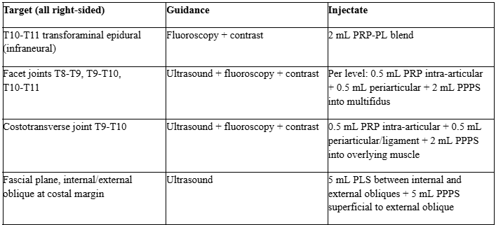
Abbreviations: PRP, platelet-rich plasma; PRP-PL, 1 mL PRP with 3 mL platelet lysate; PLS, platelet lysate solution (8 mL platelet lysate, 5% dextrose, 0.1% ropivacaine); PPPS, platelet-poor plasma solution (13 mL platelet-poor plasma, 5% dextrose, 0.25% ropivacaine).
Table 4. Biologic sample analysis
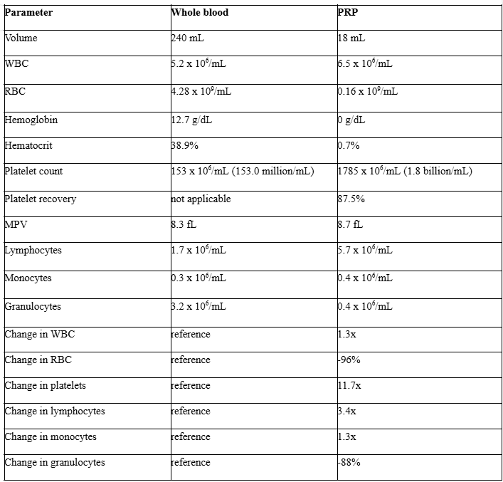
Abbreviations: MPV, mean platelet volume; PRP, platelet-rich plasma; RBC, red blood cells; WBC, white blood cells.
Table 5. Standardized Patient Registry Surveys
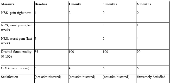
Figure 1 & 2
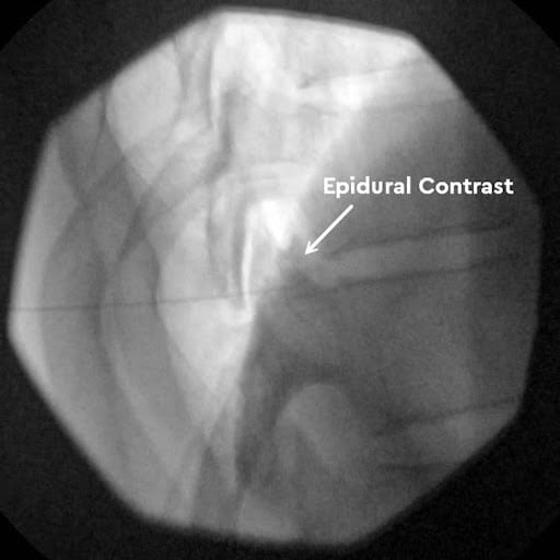
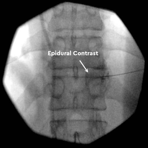
Figure 3
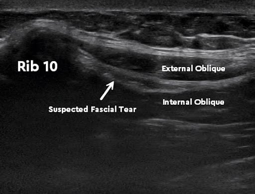
Figure 4 & 5
Corresponding author: Drew Timmermans, ND, RMSK; Regenerative Performance, Gilbert, AZ; drtimmermans@regenerativeperformance.com; 480-508-4226.
Conflicts of interest: The evaluation and treatment described were performed at the author’s own practice, and the patient paid for care directly. The author has no industry relationships relevant to this report.
Funding: None.
Informed consent: Written informed consent for publication of this de-identified case report, including the de-identified images, was obtained from the patient.