
Andrea Sulyok, ND
Can patients with Hashimoto’s thyroiditis experience persistent cognitive symptoms despite normal thyroid hormone levels? This article examines the potential roles of thyroid antibodies, tissue-level T3 activity, and objective brain performance testing in understanding brain fog beyond the standard thyroid panel.
A normal TSH and free thyroid hormone panel may not fully explain persistent brain fog in some patients with Hashimoto’s thyroiditis. Using a case example alongside current research, this article explores how thyroid autoimmunity, tissue-level T3 physiology, and objective cognitive testing may provide additional insight into persistent cognitive symptoms while identifying important directions for future research.
Thyroid dysfunction has a well-known symptom list; fatigue, weight change, hair thinning, cold intolerance, mood disturbance, and brain fog. What’s rarely discussed is what “brain fog” consists of: how quickly someone reacts, how easily they shift between competing tasks, how much effort it takes to execute under mental load. These measures sit at the intersection of thyroid physiology and brain function, where a technically normal hormone panel can coexist with active antibody elevation and suboptimal brain performance.
In Hashimoto’s thyroiditis, a real example of thyroid labs reading as controlled despite elevated antibody trends, paired with objective brain performance data, opens a discussion of what these measures may reveal about the relationship between thyroid status and brain function, from T3’s direct role in neuronal energy metabolism to the emerging evidence that thyroid antibodies themselves may act on the brain independent of hormone levels.
Objective Findings
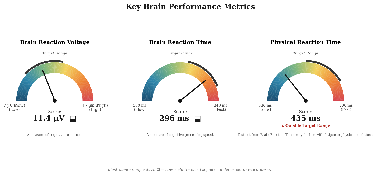
In a 54-year-old male Hashimoto patient on a stable levothyroxine dose, thyroid and metabolic labs, along with P300 brain performance testing (both obtained in December 2025), provide insight on hormone status, antibody status, and objective brain performance (Table 1, Table 2, Figure 1).
Table 1. Thyroid, Antibody, and Metabolic Labs, December 2025 (Age 54, Male)
Reflects thyroid hormone status, thyroid autoantibody titers, and associated metabolic and lipid markers from the same draw.
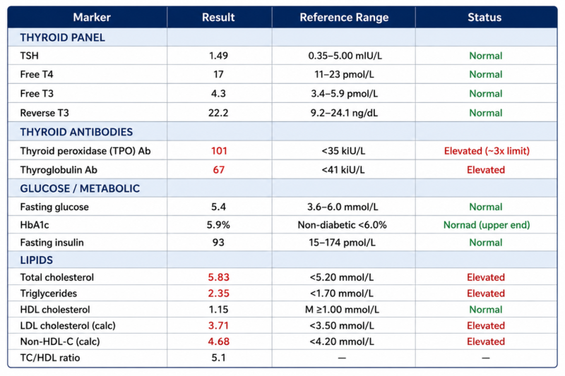
Table 2. P300 Brain Performance Findings (Baseline, Age 54)
Reflects objective brain performance testing obtained the day following the labs in Table 1.
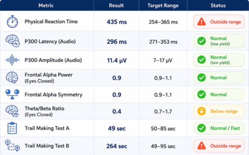
Figure 1. Key brain performance metrics, taken December 2025. Brain Reaction Voltage and Brain Reaction Time were within target range; Physical Reaction Time was not.
Interpretation
Upon inspection, TSH, Free T4, and Free T3 all read within range, and there were no classic hypothyroid symptoms; no significant weight change, no cold intolerance. By every conventional measure, the thyroid looked controlled.
The exception was thyroid antibodies, which remained persistently elevated. This pattern is consistent with what’s been described elsewhere: even after patients with Hashimoto’s thyroiditis normalize TSH and thyroid hormone levels, antibodies can remain elevated, since hormone replacement corrects the deficiency but does not eliminate the underlying autoimmune process.1
Despite the absence of hypothyroid symptoms, persistent brain fog was what prompted P300 testing in the first place. Based on the P300 scan, it may offer some insight that the brain processes normally but executes slowly. P300 latency (296 ms) and amplitude (11.4 µV) were both normal, showing no general problem with brain processing. Physical reaction time (435 ms), a measure of the neural-to-motor signal pathway, was markedly outside target range.
Two separate mechanisms could be contributing to the findings: cellular-level T3 utilization and antibody activity operating independently of the hormone markers.
Thyroid hormone, specifically intracellular T3, supports axonal conduction velocity, mitochondrial ATP production, and neuromuscular transmission.2 A noticeable pattern appeared on Trail Making testing that measures how quickly someone connects a series of dots in order, tracking visual sequencing and mental flexibility. Part A (49 sec) was fast, while Part B (264 sec), which requires alternating between numbers and letters instead of one sequence as in Part A, was markedly slow. This demands the ability to jump between two different mental tasks at once, a more complex task. Both findings point the same direction: input and evaluation appear intact, but output, motor execution and rapid task-switching were not. This is consistent with a brain that has adequate T4 but is not necessarily converting or utilizing T3 efficiently at the tissue level, a possibility discussed further.
Free T3 read within range on this panel; however, a normal serum value doesn’t necessarily mean tissue-level availability was sufficient, since T3 has to act inside cells to support nerve conduction speed, cellular energy production, and muscle signaling. Whether enough of it was reaching tissue at that level is a separate question from whether numerically labs appear in healthy ranges.
Further demonstrated with lab data: thyroid peroxidase antibodies were roughly three times the upper reference limit alongside a normal hormone panel. Proposed mechanisms for how TPO antibodies might affect brain function include direct binding to brain tissue, immune-driven neuroinflammation, and resulting disruption of neurotransmitter signaling, independent of hormone status.3
This is a single lab draw, not a trend, so neither explanation can be confirmed from this data alone.
T3 Availability and Neuronal Energy Metabolism
The brain is one of the most metabolically demanding organs in the body, and T3 plays a direct role in meeting that demand. T3 supports mitochondrial ATP production, the energy that neurons rely on for everything from maintaining resting membrane potential to firing action potentials. When T3 does not reach cells in necessary amounts, the brain’s energy supply is affected directly and this gap may show up as something like slower motor execution.
Imaging data may also support this link directly. A PET study measuring cerebral blood flow and glucose metabolism in hypothyroid adults found both were significantly reduced compared to healthy controls.4 Glucose metabolism is how cells convert fuel into ATP, and T3 regulates mitochondrial ATP production. Hence, reduced glucose metabolism on a scan is a close picture of the same outcome: T3 not adequately supporting the ATP production the brain needs to run on.
T3 also plays a role in vascular tone and cardiac output, both of which influence how much blood, oxygen and glucose actually reaches the brain tissue. Together, the two findings describe a brain that may be receiving less fuel and converting less of what it does receive, in patients with confirmed hypothyroidism. Whether this same reduction in cerebral energy metabolism can occur in patients whose Free T3 falls within reference range has not been established. However, the underlying physiology – T3’s role in mitochondrial regulation and cerebral energy metabolism – offers a plausible framework for understanding why brain performance, brain fog symptoms, and standard thyroid labs may not always align.
The Role of Thyroid Antibodies
There is growing evidence in research suggesting thyroid autoimmunity can affect the brain through pathways separate from hormone status entirely. Two studies make this case directly, both conducted in euthyroid Hashimoto’s patients, the same hormone profile discussed throughout this piece.
In one study, researchers compared two groups of patients, both with normal thyroid hormone levels at the time of testing: one group had Hashimoto’s thyroiditis (autoimmune, with elevated thyroid antibodies), and the other had thyroid diseases of non-autoimmune origin. On standardized tests of executive function and memory, the two groups didn’t differ significantly. But on one test measuring attention and response inhibition, significantly more Hashimoto’s patients scored below the normal range, and within the Hashimoto’s group, higher TPO antibody levels were related to below-normal scores.3 Both groups’ hormone levels were normal and matched, so it was proposed that any cognitive differences between them could not be explained by thyroid hormone status, only by the presence or absence of autoimmunity. A few mechanisms have been proposed for why antibodies specifically might do this: TPO antibodies binding directly to human cerebellar astrocytes, separate antibodies against central nervous system tissue that may disrupt myelin, and antibody-linked increases in inflammatory cytokines capable of altering serotonin, dopamine, and glutamate activity.3
Further, an imaging study comparing euthyroid Hashimoto’s patients to healthy controls, using event-related potentials and magnetic resonance spectroscopy, found the Hashimoto’s group’s brains responded to sounds slower and less strongly, despite normal thyroid hormone levels. Patients with the most chemical signs of neuronal stress and inflammation in the posterior cingulate gyrus were also the ones with the slowest brain wave timing.5
Both studies describe the same pattern discussed throughout this piece: a normal hormone panel sitting alongside meaningfully elevated antibodies. This reinforces antibody status as a measure worth tracking on its own, given its association with worse cognitive performance in the research above.
Conclusion
The central discussion here is that a normal thyroid panel may not guarantee optimal brain performance in those with autoimmune Hashimoto’s. Two mechanisms are consistent with this pattern: incomplete tissue-level T3 utilization and antibody activity operating independently of hormone status, though neither can be confirmed based on current data. The broader implication is that in autoimmune Hashimoto’s, a thyroid panel that reads as controlled shouldn’t be the end of the conversation when cognitive symptoms persist.
Future Directions
The mechanisms discussed here are hypotheses, not established findings. Testing them would require a few concrete steps, outlined below.
A repeat P300 assessment alongside repeat thyroid labs: hormones and both antibodies at 3 to 6 months would allow an actual trend rather than a single snapshot, and would show whether physical reaction time and Trail Making Part B shift alongside any change in antibody titer or T3 status.
If antibody activity is contributing to the brain findings discussed above, lowering levels would be the logical target to test. Selenium has the strongest evidence for doing so in autoimmune thyroiditis. A placebo-controlled RCT found selenium supplementation reduced TPO antibodies to 63.6% of baseline over three months, with no significant change in placebo or in either group’s TSH and thyroid hormone levels, indicating an antibody-specific effect.6 A subsequent meta-analysis then confirmed this across multiple trials.7 A selenium protocol followed by repeat P300 testing would show whether that antibody reduction tracks with any corresponding change in brain performance.
Ultimately, a single data point cannot establish causation between antibody status, tissue-level T3 utilization, and brain performance. A longitudinal cohort of euthyroid Hashimoto’s patients, with and without elevated antibodies, followed with paired labs and P300 testing, would be the design capable of testing whether either mechanism holds up across patients rather than in isolation.
Disclaimer
The content of this article is intended for educational purposes only and reflects a synthesis of published research and physiologic reasoning. It does not constitute medical advice, a treatment protocol, or a clinical recommendation for any individual patient. P300 testing is not a validated diagnostic tool for thyroid-related brain dysfunction and should not be interpreted as such; its use throughout this article is exploratory and hypothesis-generating only.

Author Bio
Andrea Sulyok, ND is a licensed naturopathic doctor (ND) in practice in Toronto, Canada, with a clinical interest in autoimmune health. She frequently supports patients with thyroid conditions such as Hashimoto’s thyroiditis and Graves’ disease, along with rheumatoid arthritis, lupus, and other autoimmune presentations. Her practice also emphasizes gut health, addressing conditions like IBS and dysbiosis, alongside a growing interest in brain health, including cognitive function and neuroinflammation. Andrea integrates evidence-informed nutritional, botanical, and lifestyle therapeutics to identify root causes and support sustainable, long-term wellness.
@dr.andi.the.nd
References
- Liu J, Chen Z, Liu M, Jia Y, Yao Z, Wang G. Levothyroxine replacement alleviates thyroid destruction in hypothyroid patients with autoimmune thyroiditis: evidence from a thyroid MRI study. Front Endocrinol (Lausanne). 2019;10:138.
- Bernal J. Thyroid hormones in brain development and function. In: Feingold KR, Anawalt B, Boyce A, et al, eds. Endotext. South Dartmouth, MA: MDText.com; 2015.
- Leyhe T, Müssig K. Cognitive and affective dysfunctions in autoimmune thyroiditis. Brain Behav Immun. 2014;41:261-266.
- Constant EL, de Volder AG, Ivanoiu A, Bol A, Labar D, Seghers A, Cosnard G, Melin J, Daumerie C. Cerebral blood flow and glucose metabolism in hypothyroidism: a positron emission tomography study. J Clin Endocrinol Metab. 2001;86(8):3864-3870.
- Waliszewska-Prosół M, Bladowska J, Budrewicz S, Sąsiadek M, Dziadkowiak E, Ejma M. The evaluation of Hashimoto’s thyroiditis with event-related potentials and magnetic resonance spectroscopy and its relation to cognitive function. Sci Rep. 2021;11:2480.
- Gärtner R, Gasnier BC, Dietrich JW, Krebs B, Angstwurm MW. Selenium supplementation in patients with autoimmune thyroiditis decreases thyroid peroxidase antibodies concentrations. J Clin Endocrinol Metab. 2002;87(4):1687-1691.
- Wichman J, Winther KH, Bonnema SJ, Hegedüs L. Selenium supplementation significantly reduces thyroid autoantibody levels in patients with chronic autoimmune thyroiditis: a systematic review and meta-analysis. Thyroid. 2016;26(12):1681-1692.