

A...
Chemotherapy-Induced Peripheral Neuropathy: An Evidence-Based Guide for Naturopathic Physicians
read more

A case of high-grade serous ovarian carcinoma demonstrates how serial liquid biopsy can track treatment response and progression, clarify ambiguous molecular findings, and provide a shared objective measure across naturopathic and conventional oncology care.
Circulating tumor DNA (ctDNA) testing is increasingly ordered outside the treating oncology practice, with results often reaching the naturopathic physician first. Serial liquid biopsies, drawn from multiple platforms, give integrative practitioners an objective measure that clinicians can interpret together.
This report follows a 66-year-old woman with FIGO IIIC high-grade serous ovarian carcinoma who declined first-line chemotherapy for a self-directed regimen of repurposed pharmaceuticals and dietary supplements. Serial ctDNA across three draws, read through multiple concurrent platforms, shaped her care: it documented tumor burden on her regimen, prompted her to accept conventional treatment, clarified ambiguous CHEK2/ATM findings, and tracked progression that led to a therapy change. CA-125 tracked with ctDNA throughout, while ctDNA added molecular detail no single marker could. This case offers a practical model for reading multiple ctDNA platforms alongside conventional oncology.
High-grade serous ovarian carcinoma (HGSOC) is the most common form of epithelial ovarian cancer and usually presents at an advanced stage.1,2 TP53 alterations are nearly universal; other common findings include BRCA1/2 variants, homologous recombination deficiency, and mutations in RAD51C, RAD51D, and PALB2.1,2.
Plasma ctDNA has evolved from a research tool to a routine test, often read through more than one panel — sequence-based, methylation-based, or functional — on the same patient. Serial tumor fraction correlates with CA-125 and radiologic findings and can predict recurrence.3 Naturopathic physicians now frequently review these reports, occasionally before oncology does — serving as an objective metric discussed with the patient.
Integrative oncology is ideally practiced in conjunction with standard oncology.4 This report follows one patient across three ctDNA draws and three decisions: adding chemotherapy after she initially declined it, resolving an ambiguous clonal hematopoiesis finding through multiple concurrent platforms, and switching therapy as her disease progressed.
The patient underwent biopsy in January 2025; specimens from the peritoneum and omentum showed high-grade serous carcinoma consistent with tubo-ovarian origin — primary high-grade serous adenocarcinoma of the ovary with peritoneal carcinomatosis, FIGO IIIC.
She presented for naturopathic care later that month, cachectic but alert and cognitively intact, with severe constipation attributed to tumor burden, abdominal pain, anorexia, and fatigue. BMI at admission was 18.3 kg/m² and declining; bowel dysfunction was later assessed surgically as near-obstruction.
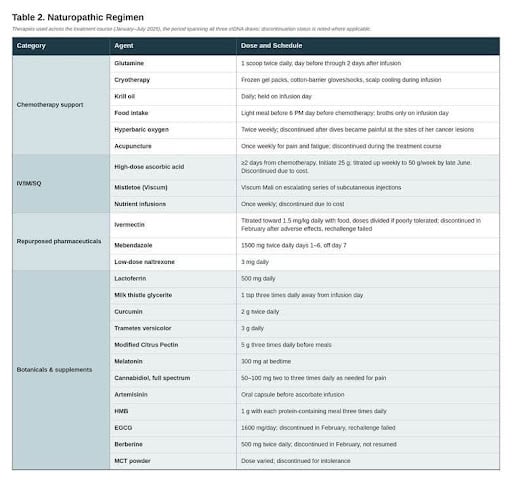
First-line carboplatin and paclitaxel had been recommended. She declined, stating that conventional treatment was inconceivable until her body was healthier, and her preference for alternative over conventional care held even as her presentation edged toward life-threatening. She had already begun a self-directed regimen of repurposed pharmaceuticals — ivermectin and fenbendazole, from a publicly circulated protocol — before the biopsy resulted and declined to stop them. I incorporated them into a supervised regimen with defined dosing, hepatic support, and monitoring; compounded mebendazole replaced fenbendazole due to its safer, human-studied hepatic profile.5-8 My role throughout was to guide treatment using evidence-based medicine and testing validated for her diagnosis. The regimen used throughout the treatment course, including agents later discontinued, is shown in Table 2.
“No single report is definitive: the April reports alone suggested near-complete response, while the July report alone would have missed that a response occurred at all. Only the full series revealed both response and progression.”
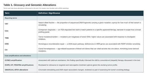
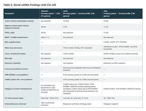
Her primary ctDNA panel — run as a 74-gene CDx — drawn in late January found a dominant TP53 L111R clone at 38.3% variant allele fraction (Table 1) with high-level BRAF and CCND1 amplifications and substantial circulating tumor burden, as measured while she pursued the self-directed regimen alone (Table 3).
I counseled her directly that her disease was too advanced for non-conventional therapy alone. I had made that case before, without effect; what changed it was her first molecular results. Presented with a quantitative measure of her tumor burden, she agreed to add conventional treatment, beginning metronomic chemotherapy in early March, about eight weeks after diagnosis.
Neither element was sufficient alone: counseling supplied clinical judgment and urgency; ctDNA supplied an external measure no clinician could be accused of interpreting self-servingly. For a patient who distrusts recommendations but accepts data — fitting many patients naturopathic physicians see — this combination may accomplish what argument alone cannot.
Two points stand out: she agreed to add chemotherapy rather than replace her regimen, and a functional drug-sensitivity assay exposed live tumor cells to a drug panel — carboplatin scored on par with the top-performing targeted agents (pazopanib, venetoclax, infigratinib, temsirolimus), paclitaxel slightly lower — a phenotypic view that reinforced the decision and surfaced options the ctDNA panels used did not evaluate.
That treatment interval was not benign. In February she developed visual disturbance, severe gastrointestinal symptoms, and gait instability attributed to ivermectin. Ivermectin and EGCG were stopped, along with berberine, turkey tail, and cannabidiol, to rest her system. Rechallenge of ivermectin and EGCG failed; berberine, turkey tail, and cannabidiol were resumed. Agents were selected against her diagnosis, symptomatology, and lab indices; tolerability set the limits of what could be sustained.
The April draw, after roughly seven weeks of chemotherapy, was read through three concurrent platforms (Table 3). Her primary panel, now run in a broader, 740-gene configuration, found the January clone undetectable, both amplifications cleared, and tumor fraction below 0.05%, the assay’s floor; several TP53 variants and an APC frameshift appeared below 0.5%. A second, independent panel returned the same class of findings. A third assay, measuring methylated ctDNA rather than sequencing variants, established a baseline for future comparison. Three technologies, applied to the same blood, converged on one conclusion: a substantial molecular response.
CA-125 confirmed the magnitude: greater than 6000 U/mL in early March, before chemotherapy began, falling to 36 U/mL two days after this draw and 27 U/mL the following week. Multiple independent measures agreed she had responded profoundly.
Two cautions applied. January’s narrower CDx panel and the broader April/July configuration serve different purposes, so variants absent from the earlier panel cannot be considered newly emergent. And the fall in tumor fraction tracks the March start of cytotoxic therapy, not the preceding regimen. Values for the same variant also differed modestly across platforms at this single point (ATM R3008H: 0.4% vs 0.22%; CHEK2 splice site: 0.6% vs 0.38%) — different chemistries reading the same signal, not a discrepancy to resolve.
The more consequential finding was structural rather than contradictory: both platforms flagged the same concern. Her primary panel placed the CHEK2 splice-site variant and ATM R3008H under “Variants of Potential Clonal Hematopoiesis,” with no therapy annotation. The second panel listed the same two variants in its main table, with annotations for olaparib and talazoparib. Still, each carried a footnote stating that the variant may be non-tumor in origin. Clonal hematopoiesis becomes more common with age and prior cytotoxic exposure, and many such variants in advanced-cancer liquid biopsies prove hematopoietic rather than tumor-derived.9 A clinician reading only the summary table, without the footnote behind it, could reasonably raise PARP eligibility prematurely.
By late July the picture had reversed. Tumor fraction rose from under 0.05% to 10.9%, and the original TP53 L111R clone returned at 17.6%. Four new amplifications appeared — CCNE1, EGFR, KIT, and PDGFRA — alongside truncating alterations in SMARCA4 and ATRX and two new ATM frameshifts (Table 1). Tumor mutational burden rose from 13.05 to 18.98 mut/Mb, crossing the assay’s threshold for likely checkpoint-inhibitor eligibility (16 mut/Mb, corresponding to a tissue TMB of 10), and genomic instability was detected. CCNE1 amplification, linked to platinum resistance, informed the switch in conventional therapy; the remaining findings — EGFR, KIT, PDGFRA, SMARCA4, and ATRX (Table 3) — informed the concurrent naturopathic regimen.
The CHEK2 splice-site variant now appeared in her primary panel’s main table with PARP-inhibitor annotations, no longer set apart as potential clonal hematopoiesis; CHEK2 R181C and ATM R3008H remained flagged. The reclassification was not arbitrary — the algorithm weighs a variant against tumor fraction, and by July that fraction had risen to 10.9% from under 0.05%, giving far more tumor-derived context. A gene name alone is not a finding, and a clonal hematopoiesis classification is not fixed to a variant; both depend on tumor burden at the moment of the draw.
CA-125 corroborated with molecular progression. After three transfusions for treatment-related toxicity, chemotherapy was held for five weeks; CA-125 rose from its nadir of 27 U/mL to 91 in early June and 1395 by July, and she was referred urgently for a palpable abdominal mass — an obvious confounder to weigh alongside the biology.
One observation is worth isolating: the low-level TP53 and APC variants detectable in April appeared while CA-125 sat at its nadir. Whether their emergence anticipated subsequent progression cannot be established from a single case, but it is the kind of early signal serial ctDNA is proposed to provide.3 The July ctDNA findings were promptly shared with her gynecologic oncologist as results indicated carboplatin and paclitaxel were no longer effective, and her cancer would likely respond to a PARP inhibitor. Her regimen was eventually switched to olaparib after 3 more rounds of carboplatin/paclitaxel had no effect.
Both clinical presentation and molecular findings guided naturopathic care. The regimen was customized to her ctDNA findings; the reported markers were actionable, using evidence-based naturopathic oncology protocols drawn from published, peer-reviewed data.10-16 Severe anorexia and a BMI of 18.3 led to a nutritional plan emphasizing calorie- and protein-dense nutrition in small portions. Chemotherapy support, IV/IM/SQ therapies, repurposed pharmaceuticals, and botanicals used across the treatment course — including agents later discontinued for cost or intolerance — appear in Table 2. The patient also opted to include a modified Joe Tippens protocol, further refined by Dr. William Makis, MD.
Serial ctDNA served as common ground for both clinician and patient, informing three key decisions and bridging a gap common in integrative practice. Each platform here — sequence-based panels of varying scope, a methylation-based assay, and a functional drug-response assay — offered a distinct view of the same tumor biology; no single view was sufficient. That is the case for ordering more than one ctDNA panel: not a winner to pick, but complementary views of the same disease.
No single report is definitive: the April reports alone suggested near-complete response, while the July report alone would have missed that a response occurred at all. Only the full series revealed both response and progression, relative to the start of cytotoxic therapy. ctDNA and CA-125 were concordant at every shared timepoint; ctDNA added molecular composition — which clones cleared, what variants emerged, and which mattered when a targeted therapy became relevant.
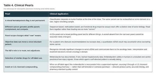
Report architecture, not disagreement between laboratories, was the issue in April: both platforms flagged the CHEK2 and ATM variants as potentially blood-derived, one by exclusion, the other by caveat. By July, independent evidence supported the PARP inhibitor that had already been selected. Clinicians should read past the summary table and raise flagged variants with oncology when a therapy decision may depend on them. Key takeaways appear in Table 4.
Serial ctDNA provided this patient’s care with a continuous trajectory and a common metric across both clinical traditions. It contributed to her decision to add conventional therapy, characterized her disease when targeted options became available, and tracked progression that prompted a regimen change. While CA-125 remained a consistent marker, ctDNA offered molecular detail CA-125 could not. For naturopathic physicians, the value lies not in interpreting these reports independently, but in recognizing important changes and ensuring findings reach the oncology team in proper context. Precision medicine is an expanding field, with genomic testing advancing every year; this case shows its practical value — concrete, validated evidence capable of moving a once-reluctant patient toward the care best suited to her scenario.