

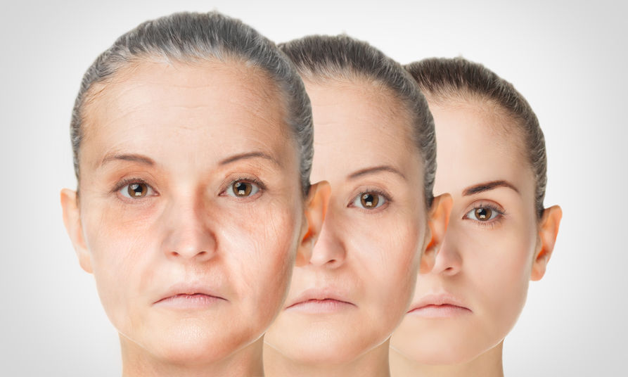
The Skin: An Outer Reflection of Inner Aging
CHRIS D. MELETIS, ND
The human skin is the largest organ of the body. It comprises 15% of body weight and has an average surface area of around 2 m2 (21.5 square feet).1 The skin has a high turnover rate, with epidermal skin cells replacing themselves approximately once every 27 days.2 The skin contains and constrains the trillions of other cells that constitute the “us” that resides within. The skin is our interface between our internal and external environment. It is highly complex, with several layers of tissues, each different in their cellular composition. Skin provides important functions, including prevention of water loss and acting as a barrier against harmful substances. Unfortunately, more than 100 million people, one-third of the US population, have at least 1 skin disease.3
Contrary to the usual advice to not judge a book by its cover, the appearance of the skin can actually reveal the inner health of a patient. The question is how do we as clinicians employ this fact as the proverbial reading of the tea leaves of what the past, present, and future may hold for our patients? Our clinically trained eye and our subconscious inner voice can discern how Patient X, Patient Y, or Patient Z feels based upon how they look during their visit. If they look unwell, that might guide our treatment. In contrast, if a patient has a radiant glow, sparkle in their eye, and exude vibrant health, we have an inkling that all is well. Even young children can look across a room and proclaim in their innocence that Aunt Betty or Uncle George doesn’t look well. Interestingly, skin blood perfusion and oxygenation impact the way we perceive a person’s health and can even influence the way people choose their mates.4 However, as clinicians, we look for specific aspects of a patient’s appearance (see Table 1) that can provide clues as to the state of their health.
Table 1: How the Skin Talks
| Condition of Skin | Possible Meaning |
| Tenting | Dehydration |
| Pallor | Anemia; impaired circulation; sympathetic/parasympathetic imbalances, etc |
| Jaundice | Liver disorder; hemolytic anemia, etc |
| Flushing | Vascular, neurological, or endocrine imbalances |
| Rashes/Dermatitis/Eczema | Internal and external altered homeostasis; nutritional imbalances; gastrointestinal dysfunction; immunological disorder, including viral, spirochete, or bacterial infection, or allergy |
| Dermatitis herpetiformis | Celiac disease, etc |
| Psoriasis | Polyamine, cytokine, or immunological imbalances; liver disorder, etc |
| Hives/Urticaria | Internal and external altered homeostasis; immunological over-reactivity; hematological disorder; mastocytosis; carcinoid syndrome |
| Dry/Moist/Oily skin | Dehydration; imbalanced essential fatty acid intake; impaired circulation; thyroid or gonadal dysfunction |
| Thin skin | Senescence; low growth hormone; high cortisol; thyroid dysfunction; vascular insufficiency, etc |
| Acne | Endocrine, biochemical, or skin microbiome imbalances |
| Rosacea | Helicobacter pylori infection; hypochlorhydria, etc |
It is not a foreign concept to clinicians to use cellular lifespan measurements to determine nutritional and wellness status. For example, we use the lifespan of red blood cells (RBCs), which live some 90-120 days, to glean insight into our patients’ health. In addition, by measuring glycosylated hemoglobin (now designated HA1c) and mean corpuscular volume (MCV), we can discern trends towards microcytic anemia (most commonly iron deficiency) or megaloblastic anemia (which frequently reflects a deficit in B12 and/or folate). Further insights are possible with measurements of RBC zinc and RBC magnesium. Our skin is nothing more than a collection of cells such as fibroblasts and keratinocytes. Therefore, it is equally important that we support a healthy lifespan of skin cells by addressing the factors mentioned in this article. In addition to discussing these primary factors that affect the aging skin and offering recommendations, I’ll delve into the connection between the health of the skin and the health of the internal body.
Skin & the Mitochondria
Addressing the health of the mitochondria is an often-overlooked way of improving aging skin. It is also a way of building a stronger foundational wellness that may result in outward changes that reflect inner health. It is now thought that mitochondria are important modulators of skin physiology.5 For example, mitochondria oversee keratinocyte differentiation via production of reactive oxygen species (ROS), which are needed for epidermal differentiation and hair follicle development.6 Mitochondria are also involved in the function and pigmentation of melanocytes.5 Further support for the mitochondria’s involvement in skin health lies in the fact that 10% of patients who have primary mitochondrial conditions exhibit skin manifestations including rashes, pigmentation abnormalities, and blueness of the extremities (acrocyanosis).5 Moreover, several skin disorders are associated with changes in mitochondrial energy metabolism, including Dupuytren’s disease, atopic dermatitis, and melanoma.5 Per Feichtinger and colleagues, “Thus, we assume that mitochondrial involvement is the rule rather than the exception in skin diseases.”5
Ultraviolet-B (UVB) radiation causes mitochondrial dysfunction and may be the primary mechanism by which long-term sun exposure can age the skin.7 Supporting mitochondrial health can therefore be beneficial for aged skin and in certain skin conditions. Mitochondrial rejuvenators of interest include coenzyme Q10 (CoQ10),8 alpha-lipoic acid,9 and resveratrol.10,11 CoQ10 and alpha-lipoic acid are used both topically and orally for skin health.
Lipofuscin, Telomeres, & the Skin Microbiome
Lipofuscin & Age Spots
Lipofuscin is a pigment composed of lipids, proteins, and carbohydrates, and it increases with age. It is produced by the synthesis of free radicals, which generate waste products, including lipofuscin.12 This age pigment is not just an indicator of aging; it has also been shown in animal studies to interfere with critical chemical and biological pathways that cells need in order to function effectively.12 Mitochondrial dysfunction can cause cells to age faster and to build up lipofuscin.13
Lipofuscin accumulation in the skin is associated with dark, smooth, brown spots known as age spots or liver spots. They are most common on the tops of the hands, but also occur on the chest and cheeks of the face. These should not be confused with the UV-induced pigmentation caused by another pigment, melanin. However, lipofuscin is not only found in the skin; it can also accumulate in neurons, cardiac myocytes, retinal epithelial pigment cells, and hepatocytes.14 Therefore, age spots on the hands or face should serve as a red flag for other conditions, including age-related macular degeneration and neurodegenerative disorders such as Alzheimer’s and Parkinson’s disease.14 In one study, an antioxidant supplement containing selenium as L-selenomethionine (300 µg), zinc (45 mg), vitamin C (270 mg), vitamin A (2.7 mg), vitamin B6 (6 mg), and vitamin E (465 mg) was given to elderly people and caused a significant drop in RBC lipofuscin.15
Telomeres
The accumulation of lipofuscin is not the only culprit in the aging process. Lipofuscin combines with other factors, including the shortening of telomeres, the protective “caps” on the ends of chromosomes that slow their degradation. A certain amount of telomere shortening is necessary to prevent cells from becoming cancerous. However, excessive telomere shortening, which occurs with aging, is associated with a number of health disorders and aging itself.16 The enzyme telomerase is responsible for maintaining telomere length. A deficiency of this enzyme in mouse models was associated with telomere shortening and skin aging.16
In humans, evidence of the connection between telomere shortening and skin health is found in a disease called dyskeratosis congenita (DC), which is associated with mutations in the gene that codes for telomerase.17 Patients with this disease often have reticulated skin pigmentation.18 People with DC have short telomeres, indicating that skin disorders that occur in these patients can be reduced by supporting telomere length.17
There is other evidence supporting the relationship between telomeres and skin health. Studies of human skin keratinocytes have found that regulation of telomere length and telomerase activity is important in these cells.16 Telomere shortening in fibroblast cells of the dermis is associated with epidermal damage.16 Since the skin acts as an environmental barrier for the body, it is exposed to ROS, which are involved in telomere shortening and aging skin.16 In part because of their exposure to ROS, telomeres in skin cells may be especially vulnerable to accelerated shortening.16 With excessive sun exposure, UV radiation damages DNA and increases telomere shortening.19 In fact, skin aging and sun damage share a common pathway that involves impaired telomeres.19 As telomere length shortens, lipofuscin also increases.14
Natural strategies for supporting telomere health include supplementing with an isolated extract of Astragalus membranaceus20 as well as resveratrol21 and other antioxidants that inhibit overproduction of ROS.
Skin Microbiome
The saying “cleanliness is next to godliness” may not apply to the skin. It’s possible to be too clean. Although in clinical practice we often acknowledge the gastrointestinal microbiome, oral microbiome, and the female reproductive microbiome, we often neglect to think about the skin microbiome.22 This is understandable, as research into the skin microbiome only began relatively recently.22 Frequent washing has been shown to alter the skin microbiota.22 Furthermore, the moment our patients place any topical substance, natural or synthetic, on their skin, they are at risk of altering their skin microecology and thus the diversity or pathogenicity of their dermal microbes. Even a natural substance can alter the skin microbiome in a deleterious fashion, either acutely or chronically. What is strong enough to help may be strong enough to harm. For example, I have witnessed many overzealous patients reporting back on the caustic effects of full-strength tea tree oil on sensitive skin.
Wound Healing & Tissue Perfusion
It’s also important to remember that our skin must breathe. Consequently, even a simple barrier of a topical agent can alter oxygenation of tissues and compound the effects of already-poor capillary oxygen delivery in some patients. In patients with cardiac issues who have decreased perfusion or in people with conditions such as peripheral artery disease, the ability of the skin to breathe becomes even more important.
Blood flow perfuses the body with oxygen, nutrients, hormones, cells, products necessary for wound healing, and platelets.23 As an example of how inner health is linked with outward appearance, blood flow to the skin plays an important role in wound healing. Individuals with heart disease or diabetes mellitus have reduced microvascular perfusion, resulting in slower wound healing.24 Researchers believe that because aged-garlic extract increased microcirculation in patients with an increased risk for cardiovascular events, it may also improve wound healing.24
Since the early 1990s, I have observed that something as simple as using an oxygen tent with a simple oxygen generator can help tissue healing when properly employed, while at the same time identifying and removing the obstacles to cure. Oxygen is not only essential to wound healing; it also guards against infection.25 Oxygen’s role in wound healing is a vast and complex topic. More information can be found in my free ebook, Oxygen: Nature’s Gift of Life and Health, available by emailing me at: [email protected].
Nitric Oxide
Nitric oxide (NO) is a molecular messenger involved in skin health. Balanced levels of NO produced in the skin support the maintenance of the skin barrier and regulate blood flow in the microvasculature. When the skin is exposed to UV light or is wounded, higher levels of nitric oxide synthase are produced, which trigger other more complex reactions.26 After the skin is exposed to UV radiation, there is a burst of NO that helps initiate melanogenesis, erythema, and immunosuppression while at the same time protecting keratinocytes against apoptosis caused by UV radiation.26 In skin wounds, increased NO synthase activity helps send infiltrating white blood cells to the site of injury and initiates the inflammation necessary for a healing response.26 Because tissue perfusion is so important for wound healing, and because NO is a vasodilator, utilizing a nitrate-rich supplement that contains beetroot may help deliver oxygen to tissues.27
Hyaluronic Acid: A Natural Moisturizer
Moisture content in the skin also impacts skin aging and its ability to heal. Hyaluronic acid (HA) is a polysaccharide and a glycosaminoglycan that acts as a natural lubricant in the skin, between joints, and in the vitreous humour of the eye. More than half of the HA in the body is concentrated in the skin.28 HA levels fall with aging and may play a role in the increased dryness and wrinkling of aged skin. HA is important for skin hydration and wound healing. Wounded tissue that is high in HA does not form scars when healing.28 HA is found in many topical skin lotions29 and can be injected into the skin,30 but it can also be used orally. Oral supplementation with HA can have beneficial results not only in the skin, but also on joint health.31
Epigenetics & the Skin
Epigenetics refers to heritable alterations in gene expression that do not involve changes in the DNA sequence. Epigenetic mechanisms include DNA methylation, histone modification, chromatin remodeling, and RNA interference. Epigenetics serve as the rheostat determining integrity, readability, and sustainability of both the cells, themselves, and the DNA and mitochondria within. Epigenetic changes in the skin can be triggered by lifestyle factors such as psychological stress,32 poor diet,33,34 and lack of sleep,35,36 in addition to exposure to UV radiation.37
Antioxidant consumption may lead to beneficial epigenetic alterations in the skin.38,39 Dietary antioxidants that protect the skin from UV radiation damage include carotenoids and polyphenols, such as apigenin (a flavonoid occurring in numerous herbs, fruits, and vegetables), quercetin, curcumin, silymarin, proanthocyanidins, and resveratrol.40 It should also be noted that nicotinamide reduces inflammation and arrests the reduction in ATP levels that occurs after exposure to UV radiation; nicotinamide has also been shown to have other beneficial effects in cell culture, rodent, and human studies, indicating that supplementation with this nutrient may have clinical usefulness.41
Skin as a Detox Organ
The skin is the barrier protecting us from the outside world. Consequently, when exposed to toxins, the skin can become a doorway leading to full body toxicity. The skin is frequently exposed to toxins such as phthalates and parabens from plastic, personal care products, and other sources. Toxins can permeate all layers of the epidermis, the skin’s uppermost layer, and subsequently be carried away into the bloodstream by the capillaries of the dermis.42 Butylparabens are known to easily penetrate the stratum corneum layer of the skin.43 Furthermore, human research indicates that dermal absorption is an important source of phthalate toxicity.44
Even something as seemingly harmless as sunscreen may contain toxic, carcinogenic chemicals that can impact skin, cells, and hormones.45 Chemicals often found in sunscreen, such as homosalate, octisalate, avobenzone, octocrylene, octinoxate, and oxybenzone, mimic hormones and disrupt testosterone, estrogen, and thyroid pathways in human cell lines and rodent studies.46,47 Other components of sunscreen promote oxidative damage in vitro and in cultured human fibroblasts.48 The toxicity of some chemicals in sunscreen is triggered by sunlight exposure, which leads to DNA damage and overproduction of ROS, according to in-vitro studies.49,50 Choosing a sunscreen without toxic chemicals can support skin health. A good source for determining the safety of sunscreen brands is the Environmental Working Group (EWG)’s website.
Conclusion
Skin is an outward mirror of our inner cells. More often than not, the skin can serve as a real-time measure of overall health. Factors affecting the skin include mitochondrial health, lipofuscin levels, telomere length, the skin microbiome, HA concentrations, epigenetics and lifestyle factors, and oxygen perfusion. Skin cells, like other cells in our body, need sufficient antioxidants. The need for antioxidants may increase, depending on sun exposure and/or exposure to environmental toxins. Addressing all factors involved in skin health can lead to a vibrant appearance on the outside and optimal health on the inside.
References:
- Slominski AT, Zmijewski MA, Semak I, et al. Melatonin, mitochondria, and the skin. Cell Mol Life Sci. 2017;74(21):3913-3925.
- Cleveland Clinic. Skin. Last reviewed March 17, 2016. Available at: https://my.clevelandclinic.org/health/articles/10978-skin. Accessed March 9, 2021.
- Chen M, Gupta V, Anselmo AC, et al. Topical delivery of hyaluronic acid into skin using SPACE-peptide carriers. J Control Release. 2014;173:67-74.
- Stephen ID, Coetzee V, Law Smith M, Perrett DI. Skin blood perfusion and oxygenation colour affect perceived human health. PLoS One. 2009;4(4):e5083.
- Feichtinger RG, Sperl W, Bauer JW, Kofler B. Mitochondrial dysfunction: a neglected component of skin diseases. Exp Dermatol. 2014;23(9):607-614.
- Hamanaka RB, Chandel NS. Mitochondrial metabolism as a regulator of keratinocyte differentiation. Cell Logist. 2013;3(1):e25456.
- Gonzalez Maglio DH, Paz ML, Ferrari A, et al. Skin damage and mitochondrial dysfunction after acute ultraviolet B irradiation: relationship with nitric oxide production. Photodermatol Photoimmunol Photomed. 2005;21(6):311-317.
- Sharma A, Soliman GM, Al-Hajaj N, et al. Design and evaluation of multifunctional nanocarriers for selective delivery of coenzyme Q10 to mitochondria. Biomacromolecules. 2012;13(1):239-252.
- Wang L, Wu CG, Fang CQ, et al. The protective effect of α-Lipoic acid on mitochondria in the kidney of diabetic rats. Int J Clin Exp Med. 2013;6(2):90-97.
- de Ligt M, Bruls YMH, Hansen J, et al. Resveratrol improves ex vivo mitochondrial function but does not affect insulin sensitivity or brown adipose tissue in first degree relatives of patients with type 2 diabetes. Mol Metab. 2018;12:39-47.
- Pollack RM, Barzilai N, Anghel V, et al. Resveratrol Improves Vascular Function and Mitochondrial Number but Not Glucose Metabolism in Older Adults. J Gerontol A Biol Sci Med Sci. 2017;72(12):1703-1709.
- Prokopiou E, Kolovos P, Georgiou C, et al. Omega-3 fatty acids supplementation protects the retina from age-associated degeneration in aged C57BL/6J mice. BMJ Open Ophthalmol. 2019;4(1):e000326.
- Tonolli PN, Martins WK, Junqueira HC, et al. Lipofuscin in keratinocytes: Production, properties, and consequences of the photosensitization with visible light. Free Radic Biol Med. 2020;160:277-292.
- Skoczyńska A, Budzisz E, Trznadel-Grodzka E, Rotsztejn H. Melanin and lipofuscin as hallmarks of skin aging. Postepy Dermatol Alergol. 2017;34(2):97-103.
- Clausen J, Nielsen SA, Kristensen M. Biochemical and clinical effects of an antioxidative supplementation of geriatric patients. A double blind study. Biol Trace Elem Res. 1989;20(1-2):135-151.
- Buckingham EM, Klingelhutz AJ. The role of telomeres in the ageing of human skin. Exp Dermatol. 2011;20(4):297-302.
- Kirwan M, Dokal I. Dyskeratosis congenita: a genetic disorder of many faces. Clin Genet. 2008;73(2):103-112.
- Marciniak RA, Johnson FB, Guarente L. Dyskeratosis congenita, telomeres and human ageing. Trends Genet. 2000;16(5):193-195.
- Kosmadaki MG, Gilchrest BA. The role of telomeres in skin aging/photoaging. Micron. 2004;35(3):155-159.
- Salvador L, Singaravelu G, Harley CB, et al. A Natural Product Telomerase Activator Lengthens Telomeres in Humans: A Randomized, Double Blind, and Placebo Controlled Study. Rejuvenation Res. 2016;19(6):478-484.
- Gutlapalli SD, Kondapaneni V, Toulassi IA, et al. The Effects of Resveratrol on Telomeres and Post Myocardial Infarction Remodeling. Cureus. 2020;12(11):e11482.
- Dréno B, Araviiskaia E, Berardesca E, et al. Microbiome in healthy skin, update for dermatologists. J Eur Acad Dermatol Venereol. 2016;30(12):2038-2047.
- Matienzo D, Bordoni B. Anatomy, Blood Flow. In: StatPearls. Treasure Island, FL: StatPearls Publishing; 2021.
- Wlosinska M, Nilsson AC, Hlebowicz J, et al. Aged garlic extract preserves cutaneous microcirculation in patients with increased risk for cardiovascular diseases: A double-blinded placebo-controlled study. Int Wound J. 2019;16(6):1487-1493.
- Younis I. Role of oxygen in wound healing. J Wound Care. 2020;29(Sup5b):S4-s10.
- Cals-Grierson MM, Ormerod AD. Nitric oxide function in the skin. Nitric Oxide. 2004;10(4):179-193.
- Baião DDS, Silva D, Paschoalin VMF. Beetroot, a Remarkable Vegetable: Its Nitrate and Phytochemical Contents Can be Adjusted in Novel Formulations to Benefit Health and Support Cardiovascular Disease Therapies. Antioxidants (Basel). 2020;9(10).
- Manuskiatti W, Maibach HI. Hyaluronic acid and skin: wound healing and aging. Int J Dermatol. 1996;35(8):539-544.
- Sundaram H, Mackiewicz N, Burton E, et al. Pilot Comparative Study of the Topical Action of a Novel, Crosslinked Resilient Hyaluronic Acid on Skin Hydration and Barrier Function in a Dynamic, Three-Dimensional Human Explant Model. J Drugs Dermatol. 2016;15(4):434-441.
- Espinoza L, Vinshtok Y, McCreesh J, et al. Kinetic energy-assisted delivery of hyaluronic acid for skin remodeling in middle and lower face. J Cosmet Dermatol. 2020;19(9):2277-2281.
- Cicero AFG, Girolimetto N, Bentivenga C, et al. Short-Term Effect of a New Oral Sodium Hyaluronate Formulation on Knee Osteoarthritis: A Double-Blind, Randomized, Placebo-Controlled Clinical Trial. Diseases. 2020;8(3):26.
- Lee CM, Watson REB, Kleyn CE. The impact of perceived stress on skin ageing. J Eur Acad Dermatol Venereol. 2020;34(1):54-58.
- Roszkiewicz M, Dopytalska K, Szymańska E, et al. Environmental risk factors and epigenetic alternations in psoriasis. Ann Agric Environ Med. 2020;27(3):335-342.
- Orioli D, Dellambra E. Epigenetic Regulation of Skin Cells in Natural Aging and Premature Aging Diseases. Cells. 2018;7(12).
- Ramos-Lopez O, Milagro FI, Riezu-Boj JI, Martinez JA. Epigenetic signatures underlying inflammation: an interplay of nutrition, physical activity, metabolic diseases, and environmental factors for personalized nutrition. Inflamm Res. 2021;70(1):29-49.
- Jang SI, Lee M, Han J, et al. A study of skin characteristics with long-term sleep restriction in Korean women in their 40s. Skin Res Technol. 2020;26(2):193-199.
- Lee Y, Shin MH, Kim MK, et al. Increased Histone Acetylation and Decreased Expression of Specific Histone Deacetylases in Ultraviolet-Irradiated and Intrinsically Aged Human Skin In Vivo. Int J Mol Sci. 2021;22(4):2032.
- Letsiou S, Kapazoglou A, Tsaftaris A. Transcriptional and epigenetic effects of Vitis vinifera L. leaf extract on UV-stressed human dermal fibroblasts. Mol Biol Rep. 2020;47(8):5763-5772.
- Henning SM, Wang P, Carpenter CL, Heber D. Epigenetic effects of green tea polyphenols in cancer. Epigenomics. 2013;5(6):729-741.
- Michalak M, Pierzak M, Kręcisz B, Suliga E. Bioactive Compounds for Skin Health: A Review. Nutrients. 2021;13(1):203.
- Snaidr VA, et al. Nicotinamide for photoprotection and skin cancer chemoprevention: A review of efficacy and safety. Exp Dermatol. 2019;28 Suppl 1:15-22.
- Stepanov D, Canipa S, Wolber G. HuskinDB, a database for skin permeation of xenobiotics. Sci Data. 2020;7(1):426.
- Seko N, Bando H, Lim CW, et al. Theoretical analysis of the effect of cutaneous metabolism on skin permeation of parabens based on a two-layer skin diffusion/metabolism model. Biol Pharm Bull. 1999;22(3):281-287.
- Pan W, Zeng D, Ding N, et al. Percutaneous Penetration and Metabolism of Plasticizers by Skin Cells and Its Implication in Dermal Exposure to Plasticizers by Skin Wipes. Environ Sci Technol. 2020;54(16):10181-10190.
- Al-Eitan LN, Aljamal HA, Alkhatib RQ. Gas chromatographic-mass spectrometric analysis of sunscreens and their effects on mice liver and kidney enzyme function. Clin Cosmet Investig Dermatol. 2019;12:11-21.
- Schlumpf M, Cotton B, Conscience M, et al. In vitro and in vivo estrogenicity of UV screens. Environ Health Perspect. 2001;109(3):239-244.
- Schlumpf M, Schmid P, Durrer S, et al. Endocrine activity and developmental toxicity of cosmetic UV filters–an update. Toxicology. 2004;205(1-2):113-122.
- Dunford R, Salinaro A, Cai L, et al. Chemical oxidation and DNA damage catalysed by inorganic sunscreen ingredients. FEBS Lett. 1997;418(1-2):87-90.
- Knowland J, McKenzie EA, McHugh PJ, Cridland NA. Sunlight-induced mutagenicity of a common sunscreen ingredient. FEBS Lett. 1993;324(3):309-313.
- Mei N, Hu J, Xia Q, et al. Cytotoxicity and mutagenicity of retinol with ultraviolet A irradiation in mouse lymphoma cells. Toxicol In Vitro. 2010;24(2):439-444.

Chris D. Meletis, ND is an educator, international author, and lecturer. He has authored 18 books and more than 200 scientific articles in prominent journals and magazines. Dr Meletis served as Dean of Naturopathic Medicine and CMO for 7 years at NUNM. He was recently awarded the NUNM Hall of Fame award by OANP, as well as the 2003 Physician of the Year by the AANP. Dr Meletis spearheaded the creation of 16 free natural medicine healthcare clinics in Portland, OR. Dr Meletis serves as an educational consultant for Fairhaven Health, Berkeley Life, TruGen3, US BioTek, and TruNiagen. He has practiced in Beaverton, OR, since 1992.


