

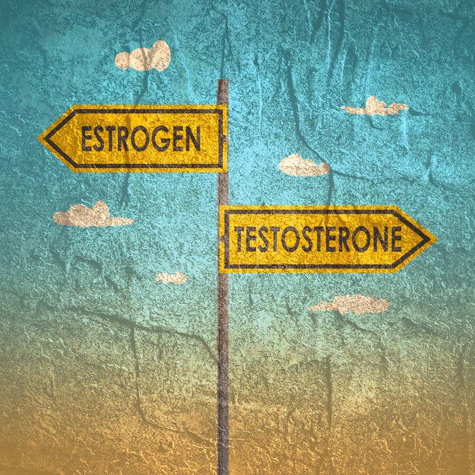
Steroidogenesis: Looking Beyond Estrogen and Testosterone
CHRIS D. MELETIS, ND
When a woman or man becomes “pausal,” whether it be menopause or andropause, low hormone levels can impact many aspects of life. Low hormone levels affect bravado, finesse, and the physiological capability needed to pivot during times of stress. Low hormone levels also weaken one’s ability to address variables that are part of living in a modern and contemporary world.
Treating hormone levels in older patients is critical. Yet, as functional medicine providers, we must be wary not to fall into reductionism when it comes to the effects of a given steroidal hormone. This reductionist trap has a long history in hormone replacement therapy. Let’s remember that not too long ago the treatment of choice following a complete hysterectomy and oophorectomy was Premarin®. Yet, the ovaries produce more than just estrogens. Although unopposed estrogen has since been shown to be potentially harmful to women, providers still treat with estrogen alone to mitigate select symptoms. So why limit treatment to 1 hormone? Is this a philosophical approach or merely chasing symptoms?
It’s important to remember men and women are more than a blend of gonadal hormones such as testosterone, estrogens, and progesterone. Many other hormones and hormonal pre-cursors influence health. It’s a big mistake to chase the end products of steroidogenesis without ensuring sufficient balance of upstream hormones and substrates.
Supporting hormonal homeostasis is challenging. We live in a world with many exogenous factors and endocrine disruptors influencing hormonal balance. Supporting upstream products like pregnenolone and DHEA can contribute to hormonal balance even when downstream hormones like estrogen and testosterone are impacted. In this article, I’ll address the importance of supporting optimal levels of hormones and their precursors, and why rejuvenating mitochondrial health is critical to ensure optimal hormone production.
Avoiding the Myopic Trap
Based on steroidal endocrinology articles, we have all been led down the primrose path, which reports that when estradiol, estriol, and testosterone are addressed, select signs and symptoms will resolve. However, this myopic approach fails to consider other important hormones like pregnenolone, DHEA, and cortisol.
One of my patients serves as a good case study demonstrating where that type of myopic approach can fall short. An 80-year-old female navy veteran complained of classical symptoms of Cushing’s disease. Yet, over the course of countless visits to an endocrinologist, internist, and other specialists, there was no definitive diagnosis. When she arrived at my office, I ordered a salivary cortisol x 4 test, which demonstrated that all the diurnal markers were literally at their upper limits of quantification. A 24-hour urinary cortisol test confirmed the results. I referred the patient for a brain MRI that revealed a pituitary adenoma. The patient had suffered from symptoms of Cushing’s disease for many years before being diagnosed.
The treatment she was offered was a full pituitary gland resection. The challenge of recovery from the pituitary resection began when the endocrinologists informed her that all she needed was 20 mg of hydrocortisone daily, in 2 divided doses. She was also prescribed an injectable growth hormone, thyroid hormone, and vasopressin to control electrolytes and water management.
The flaw in this approach was that it did not address cortisol production that occurs as a strategic trickle throughout the day. The endocrinologist also failed to address pregnenolone and DHEA, in addition to gonadal hormones. Instead, I made sure that the patient was taking cortisol to mimic “life event” cortisol production and diurnal rhythms. The body naturally makes cortisol under many stress-producing life events. A natural morning surge also occurs to mobilize blood sugar and help awaken and prepare the body for the day. I also incorporated pregnenolone and DHEA into this patient’s program, which proved critical to the healing process. Both upstream hormones serve as more than merely a step-in steroidogenesis. They have unique, currently known functions, and we can only imagine others yet to be determined.
The Origins of Steroidogenesis
To understand the importance of supporting upstream hormones, we must first look at how hormones are produced. The production of steroids begins when cholesterol is converted into pregnenolone by the CYP11A1 enzyme. This conversion occurs at the inner mitochondrial membrane.1 The mitochondria and smooth endoplasmic reticulum enzymes convert pregnenolone to testosterone, estrogen, progesterone, cortisol, or DHEA.2 This is why pregnenolone is called the “mother” (or grandmother) of all hormones.
The mitochondria play a critical role in steroidogenesis. This is why steroidogenic cells, such as adrenocortical cells in the adrenal glands, granulosa and theca cells in the ovary, Leydig cells in the testis, and syncytial trophoblast cells in the placenta, are all rich in mitochondria.3
For example, the Leydig cells in the testicles are responsible for testosterone synthesis. These cells decline with age, contributing to hypogonadism. As aging occurs, cellular changes happen in the steroidogenic enzymes of the mitochondria and smooth endoplasmic reticulum.4 These enzymatic changes impair the conversion of cholesterol to pregnenolone and pregnenolone to testosterone.
Given the mitochondria’s important role in producing steroids, any regimen designed to balance hormones should also include mitochondrial support nutrients such as coenzyme Q10 and nicotinamide riboside. Nicotinamide riboside increases levels of NAD+ by 100% and was found to restore the health of mitochondria in mouse models and human studies.5,6
Steroidogenesis can also occur in the skin,2 which isn’t surprising given that vitamin D, a hormone, is produced after exposure to sunlight. Skin cells contain all of the biochemical components needed for the synthesis of glucocorticoids, androgens, and estrogens either from precursors circulating systemically or via the conversion of cholesterol to pregnenolone. Pregnenolone is transformed to biologically active steroids such as corticosterone, cortisol, testosterone, dihydrotestosterone, and estradiol.2 Glucocorticoids, androgens, and estrogens produced in the skin affect functions of the epidermis as well as local and systemic immune activity.2
Impairment in the steroidogenic activities of the skin can result in disruptions that lead to systemic disorders, and even something as simple as sunburn from ultraviolet B radiation can affect the production of glucocorticoids.2 As Slominski et al pointed out in their 2013 research article about steroidogenesis, “The skin can be defined as an independent steroidogenic organ, whose activity can affect its functions and the development of local or systemic inflammatory or autoimmune diseases.”2
Pregnenolone
Pregnenolone is critical for many aspects of health and is the precursor to all steroid hormone production. Any hormonal support regimen should consider low levels of upstream precursor steroidal hormones, measured with lab testing. Prior to supplementing with testosterone or estrogen, measuring pregnenolone should be a strong consideration.
Pregnenolone is involved in the health of the uterus and ovaries and regulates the expression of estrogen receptors in these female reproductive organs.7 In animal research, pregnenolone decreased ovarian cell proliferation, suggesting it may suppress ovarian cancer.7 Its effects on the uterus in animal studies also suggest it may be helpful in endometriosis.7
Another potential application of pregnenolone is for pain. In a randomized, double-blind, placebo-controlled clinical trial of US military veterans with chronic low back pain, participants were given escalating pregnenolone doses of 100 mg for 1 week, 300 mg for 1 week, 500 mg for 2 weeks, or a placebo.8 Veterans in the pregnenolone group experienced clinically meaningful reductions in low back pain compared to the placebo group.
As mentioned earlier, pregnenolone is also essential in testosterone synthesis. When treating low testosterone, the upstream levels of pregnenolone, DHEA, and androstenedione should also be addressed. In addition, the impairment in mitochondrial enzymes that occurs during hypogonadism indicates mitochondrial nutrients are necessary to support testosterone production during aging.
Neurosteroids
Steroid hormones are key regulators of brain activity and behavior.9 They play critical roles in regulating physiological activities such as food intake, wakening, reproduction, and sexual behavior.9 They are also involved in the regulation of mood and memory.9 Furthermore, steroids help cope with stress and high levels or hypersensitivity to those hormones involved in reward-related behaviors that have been implicated in drug abuse.10
Some steroids are produced in the brain and are known as neurosteroids. Neurosteroids include pregnenolone and its metabolites pregnenolone-sulfate and allopregnanolone, as well as DHEA. Neurosteroids are involved in mood regulation, cognitive function, and age-related impairment.11,12 Many of the benefits of pregnenolone can be attributed to its actions as a neurosteroid.
As neurosteroids, pregnenolone and its derivatives are involved in many aspects of brain health, including enhancing learning and memory, relieving depression, improving locomotor activity, and promoting neuronal cell survival.13 In addition, pregnenolone has a calming effect. Human research over 8 weeks also indicated pregnenolone can enhance functional capacity in people with schizophrenia, though it did not improve cognitive symptoms.14
Don’t Neglect DHEA
Dehydroepiandrosterone (DHEA) is a steroid hormone produced in the adrenal cortex and in smaller amounts in the gonads. However, DHEA and its sulfated form (DHEA-S) can also be synthesized in the brain, indicating their function as neurosteroids. In this role, DHEA and DHEA-S are neuroprotective, promote neurite growth, and have anti-inflammatory, antioxidant, and anti-glucocorticoid effects.15
DHEA’s neurosteroid effects occur due to its conversion to either testosterone and dihydrotestosterone via androgen receptors or estradiol via estrogen receptors, both of which are present in most parts of the human brain.15 DHEA may also produce effects indirectly via the activities of its metabolites formed locally in the brain.15
Plasma DHEA-S levels are at their highest in the 3rd decade of life; they then gradually fall at an average rate of 1%-4% per year in men (during their 40s to 80s) and approximately 2% per year in women.16 By the onset of menopause, DHEA levels have already declined in women, on average, by 60%.17 Between women, there is a significant difference in the circulating concentrations of DHEA. Some postmenopausal women can have barely detectable serum levels, while others’ DHEA levels are normal.17
The age-related decline of DHEA-S serum levels occurs together with an increased prevalence of several disorders.16 Low DHEA levels are thought to be involved in a host of issues associated with post-menopause, including:
- Osteoporosis
- Muscle loss
- Vaginal atrophy
- Fat accumulation
- Hot flashes
- Skin atrophy
- Type 2 diabetes
- Memory loss
- Cognition loss
- Alzheimer’s disease17
Research indicates that vaginal atrophy, a problem due to the hormone deficiencies of menopause, can be quickly improved or eliminated by intravaginal administration of DHEA without systemic exposure to estrogens.18,19 DHEA-S levels also negatively correspond to ischemic heart disease, cardiovascular mortality, atherosclerosis, osteoporosis, and inflammatory diseases.16 When testing for DHEA-S, it is important to remember that plasma concentrations of DHEA-S may not correspond to levels in the central nervous system due to the innate ability of the brain to synthesize neurosteroids.20
Conclusion
It’s important to get a clear understanding of the underlying causes of patients’ symptoms by addressing downstream hormones like testosterone and estrogen, and by paying attention to the upstream precursor pregnenolone. DHEA is another downstream hormone that often takes a backseat to supplementation with testosterone or estrogen, but it is nevertheless critical for patients’ health. Taking note of patients’ cortisol concentrations and lowering levels that are too high, or increasing levels that are too low in a circadian-dependent fashion, can lead to meaningful clinical improvement. Finally, paying attention to mitochondrial health is crucial during a hormonal support regimen since mitochondria play a critical role in pregnenolone production. Before supplementing with any hormones, it’s important to quantify levels by testing, not guessing. In my clinical practice, I have also found it helpful to perform an organic acids test for environmental pollutants to determine if the patient is exposed to any environmental toxins that could be disrupting their endocrine system.
References:
- Miller WL. Steroid hormone synthesis in mitochondria. Mol Cell Endocrinol. 2013;379(1-2):62-73.
- Slominski A, Zbytek B, Nikolakis G, et al. Steroidogenesis in the skin: implications for local immune functions. J Steroid Biochem Mol Biol. 2013;137:107-123.
- Bassi G, Sidhu SK, Mishra S. The Expanding Role of Mitochondria, Autophagy and Lipophagy in Steroidogenesis. Cells. 2021;10(8):1851.
- Racchi M, Balduzzi C, Corsini E. Dehydroepiandrosterone (DHEA) and the aging brain: flipping a coin in the “fountain of youth”. CNS Drug Rev. 2003;9(1):21-40.
- Hou Y, Lautrup S, Cordonnier S, et al. NAD(+) supplementation normalizes key Alzheimer’s features and DNA damage responses in a new AD mouse model with introduced DNA repair deficiency. Proc Natl Acad Sci U S A. 2018;115(8):E1876-E1885.
- Airhart SE, Shireman LM, Risler LJ, et al. An open-label, non-randomized study of the pharmacokinetics of the nutritional supplement nicotinamide riboside (NR) and its effects on blood NAD+ levels in healthy volunteers. PLoS One. 2017;12(12):e0186459.
- Shin YY, Kang EJ, Jeong JS, et al. Pregnenolone as a potential candidate for hormone therapy for female reproductive disorders targeting ERβ. Mol Reprod Dev. 2019;86(1):109-117.
- Naylor JC, Kilts JD, Shampine LJ, et al. Effect of Pregnenolone vs Placebo on Self-reported Chronic Low Back Pain Among US Military Veterans: A Randomized Clinical Trial. JAMA Netw Open. 2020;3(3):e200287.
- Vallée M, Vitiello S, Bellocchio L, et al. Pregnenolone can protect the brain from cannabis intoxication. Science. 2014;343(6166):94-98.
- Piazza PV, Le Moal M. Glucocorticoids as a biological substrate of reward: physiological and pathophysiological implications. Brain Res Brain Res Rev. 1997;25(3):359-372.
- Baulieu EE, Robel P, Schumacher M. Neurosteroids: beginning of the story. Int Rev Neurobiol. 2001;46:1-32.
- Vallée M, Mayo W, Koob GF, Le Moal M. Neurosteroids in learning and memory processes. Int Rev Neurobiol. 2001;46:273-320.
- Weng JH, Chung BC. Nongenomic actions of neurosteroid pregnenolone and its metabolites. Steroids. 2016;111:54-59.
- Marx CE, Lee J, Subramaniam M, et al. Proof-of-concept randomized controlled trial of pregnenolone in schizophrenia. Psychopharmacology (Berl). 2014;231(17):3647-3662.
- Stárka L, Dušková M, Hill M. Dehydroepiandrosterone: a neuroactive steroid. J Steroid Biochem Mol Biol. 2015;145:254-260.
- Chen CY, Wu CC, Huang YC, Hung CF, Wang LJ. Gender differences in the relationships among neurosteroid serum levels, cognitive function, and quality of life. Neuropsychiatr Dis Treat. 2018;14:2389-2399.
- Labrie F. DHEA, important source of sex steroids in men and even more in women. Prog Brain Res. 2010;182:97-148.
- Labrie F, Bélanger A, Pelletier G, Martel C, Archer DF, Utian WH. Science of intracrinology in postmenopausal women. Menopause. 2017;24(6):702-712.
- Labrie F, Archer DF, Koltun W, et al. Efficacy of intravaginal dehydroepiandrosterone (DHEA) on moderate to severe dyspareunia and vaginal dryness, symptoms of vulvovaginal atrophy, and of the genitourinary syndrome of menopause. Menopause. 2018;25(11):1339-1353.
- Beattie MC, Adekola L, Papadopoulos V, Chen H, Zirkin BR. Leydig cell aging and hypogonadism. Exp Gerontol. 2015;68:87-91.

Chris D. Meletis, ND is an educator, international author, and lecturer. He has authored 18 books and more than 200 scientific articles in prominent journals and magazines. Dr Meletis served as Dean of Naturopathic Medicine and CMO for 7 years at NUNM. He was recently awarded the NUNM Hall of Fame award by OANP, as well as the 2003 Physician of the Year by the AANP. Dr Meletis spearheaded the creation of 16 free natural medicine healthcare clinics in Portland, OR. Dr Meletis serves as an educational consultant for Fairhaven Health, Berkeley Life, TruGen3, US BioTek, and TruNiagen. He has practiced in Beaverton, OR, since 1992.


