

Pathogenesis of Parkinson’s: Implications for Early Diagnosis and Treatment
Sonja M. Halsey
Steven Sandberg-Lewis, ND, DHANP
Parkinson’s disease (PD) is a multisystem disorder involving dopaminergic, noradrenergic, serotoninergic and cholinergic systems, and characterized by motor and non-motor symptoms. It is the second most common neurodegenerative disease after Alzheimer’s disease,1 affecting 1 million people in the United States every year.2 Both sporadic and familial forms are known.3 The disease affects 1% of the population over the age of 604 and causes progressive disability that can be slowed, but not stopped or reversed, by treatment. PD is hypothesized to be due to a combination of genetic and environmental factors, though environmental triggers are unknown.
A Brief Review
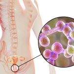
The 2 major neuropathological findings are loss of pigmentation of the substantia nigra and the presence of Lewy bodies within neurons. The degeneration of neurons in the substantia nigra leads to striatal dopamine deficiency, which is responsible for the major motor symptoms of the disease.1 Lewy bodies are neural inclusions whose main component are misfolded alpha-synuclein aggregates.5,6 These inclusions develop within specific types of projection neurons in all portions of the nervous system,7 though the “core” of the neuronal lesions is the progressive degeneration of dopamine neurons in the central nervous system (CNS), which accounts for most of its end-stage symptoms. However, it is also well known that these lesions occur outside the CNS, particularly in the enteric nervous system (ENS).1
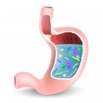
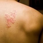
The signs and symptoms of PD are numerous, with both premotor and motor components. Premotor symptoms can include sleep disturbances, decreased sense of smell, gastrointestinal (GI) dysfunction, sexual dysfunction, depression, weakness, malaise, slow thinking, and seborrheic dermatitis.8 Among these symptoms, GI dysfunctions are the most common non-motor symptoms and worsen with progression of the disease. Some of these symptoms may include: sialorrhea (hypersalivation), swallowing disorders, dysphagia, acid regurgitation, pyrosis, early satiety, weight loss, constipation, gastric atony, and incomplete rectal emptying.9-11 In PD, dysphagia, impaired gastric emptying, and constipation may precede its clinical diagnosis for years.12 There is also strong evidence of an increased occurrence of small intestinal bacterial overgrowth (SIBO) in PD patients.13
Currently, PD is a clinical diagnosis. No laboratory biomarkers exist yet for this condition, and findings on routine MRI and CT are unremarkable.14 The 4 current cardinal diagnostic signs of PD include resting tremor, rigidity, bradykinesia, and postural instability.15 However, by this stage, premotor symptoms can be advanced. A new criterion for diagnosing PD at the premotor stage is needed. Chadhuri et al have proposed that a combined approach, with the addition of a standardized non-motor symptom scale, will aid in a more complete and early diagnosis.16
One of the main treatment options for PD is levodopa, a pro-drug of dopamine. Levodopa (L-dopa) products are formulated with aromatic amino acid decarboxylase inhibitors, such as carbidopa, to prevent the metabolism of levodopa in the GI tract and allow increased L-dopa to the brain. Food affects the absorption of levodopa, with high-protein foods competing for uptake, hence reducing effects.17 Long-term levodopa therapy is associated with motor fluctuations and dyskinesias, leading some clinicians to postpone its use.18 Levodopa/carbidopa also have a long list of side effects.19
Conventional therapies for GI symptoms are also employed in treatment. Hypersalivation is reduced by anticholinergics or botulinum toxin injections. Therapy for dysphagia may include rehabilitation, surgery, and pharmacologic treatment. Constipation is managed with laxatives and prokinetics.20-24
Lewy Body Pathogenesis – A Closer Look at the ENS
The biological process that underlies the motor and non-motor phases of sporadic PD is not simultaneous at all the susceptible nervous system sites, as seen in autopsy-based studies. Observations indicate that initial Lewy body sites include the anterior olfactory structures, portions of the ENS, and the dorsal nucleus of the vagus nerve.25-27 These vulnerable regions are interconnected anatomically, and the physical contact of these regions may play a key role in PD pathogenesis, since all of the vulnerable nerve cells are interconnected projection neurons with long and sparingly mylelinated axons.28-30 It is possible that movement of the inclusions occurs via retrograde axonal transport and transynaptic transmission of an indeterminate pathogen at the myenteric and submucosal plexus, which gains access to the lower brain via the vagal nerve, and then ascends through the medulla, and from there into the pons and midbrain until it reaches the substantia nigra.31-33
Research has shown that Lewy body lesions at the ENS occur at a very early stage of the disease, even before the involvement of the CNS. This has led to the hypothesis that the ENS could be critical in the pathophysiology of PD, as it could represent a point of entry for an environmental factor to initiate the pathological process.1 Forsyth et al conducted clinical studies in patients recently diagnosed with PD to determine if increased intestinal permeability was more prevalent in PD patients than in control subjects without the disease. All patients in the study had not yet started drug therapy for PD. The data showed that PD subjects had significantly greater intestinal permeability than controls. It was also shown that hyper-permeability significantly correlated with increased intestinal mucosal staining for E coli, nitrotyrosine, alpha-synuclein, and lipopolysaccharide (LPS) in PD subjects’ sigmoid biopsy tissue.7
Authors of this study postulated that increased gut permeability in patients with genetic susceptibility to PD may be a pivotal early step promoting a pro-inflammatory and oxidative environment that contributes to the initiation or progression of PD. Translocation of bacteria and bacterial products into the mucosa of the intestines is known to increase the oxidative stress burden on the ENS, including the phosphorylation of alpha-synuclein34 and subsequent misfolding and inclusion of these proteins.7 The Honolulu Heart Study also found that constipation was associated with the subsequent development of PD and suggests that the presence of Lewy bodies and reduced dopamine levels, observed in the myenteric plexus of patients with PD, may predate CNS manifestations.35,36
Screening High Risk and Parkinson’s Associated Risk Syndrome Patients
In 2010, researchers coined the term Parkinson’s Associated Risk Syndrome (PARS) to help determine at-risk and subclinical stages of the disease. PARS looks at genetic predisposition, neuroimaging changes, premotor signs and symptoms, and subtle prediagnostic neurologic features. The premotor stages of PARS include olfactory dysfunction, altered sleep patterns, rapid eye movement (REM) sleep behavior disorders, anxiety and depression, constipation, diminished color discrimination, and contrast sensitivity.37,38 Screening regularly for these patterns, as well as identifying high-risk patients (familial/age), could lead to earlier and more successful long-term treatment.15 Special testing may also be employed to screen for PARS. Intestinal permeability can be tested using 24-hour sucralose, lactulose, or mannitol. Intestinal antigenic or barrier screens may also be used to help assess intestinal permeability in suspected Parkinson’s patients.39 Possibly, sigmoidoscopic biopsy may reveal Lewy bodies in the submucosal and myenteric plexus of the ENS.7
Currently, grants have been funded to research differences in the microflora of the nose and gut in PD patients compared with controls. These results may have a major impact on prevention and early treatment for the gastroenterologist.40
Brush Border Healing & Diet
An important approach to treating a patient at high risk for PD or in premotor stages of the disease is to heal the gut. This can occur through diet and supplementation. The Gut and Psychology Syndrome Introductory Diet aids in healing the brush border by removing irritants such as fiber from the diet and nourishing the gut lining with amino acids, gelatin, glucosamine, fats, vitamins and minerals, and beneficial bacteria.41 Brush-border healing can also be supported by a number of supplements, including colostrum, L-glutamine, vitamins A and D, fish oil, N-acetylcysteine (NAC), turmeric, and resveratrol.39
Based on Dr Laurie Mischley’s book, Natural Therapies for Parkinson’s Disease,42 guidelines should be followed long-term to slow the progression of PD. Some tips she recommends for patients include:
- Eat a variety of organic fruits and vegetables
- Avoid dairy
- Eat meat away from medications (or amino acid therapy)
- Decrease homocysteine (elevated from levodopa) with B6, B12, and folic acid
- Increase omega-3 for its neuro-inflammatory-modulating effects
- Use turmeric liberally! It contains antioxidants and anti-inflammatory compounds. Curcumin also alleviates the effects of glutathione depletion.
- Increase magnesium. This is great for relaxation, sleep, and constipation.
Amino Acid Therapy
The very process of PD is associated with depletion of dopamine, tyrosine, tyrosine hydroxylase, norepinephrine, serotonin, and their neuroreceptors. Depletion is only exacerbated by drug administration of levodopa and carbidopa.43-47 Amino acid therapy is being talked about more in Parkinson’s communities, yet research on amino acid therapy in conjunction with, or as an alternative to, levodopa in early stage PD is very limited. Tyrosine is important for the thyroid and other neural systems in the periphery and enteric nervous system. L-dopa’s depletion of serotonin levels in turn produces tachyphylaxis of L-dopa, rendering the drug ineffectual during the depletion no matter how much is administered.45-48
In a case study report, an amino acid supplementation protocol was administered alongside conventional L-dopa administration, to assess optimal management and response to L-dopa treatment. Treatment was initiated with administration of 5-HTP and L-tyrosine, with the addition of important co-factors: vitamin C, calcium citrate, vitamin B6, folate, L-lysine, L-cysteine, and selenium. The results indicated that amino acid administration is effective alongside L-dopa, contributing to optimal dosing of the drug without adverse reactions, as well as a decrease in patient side effects.49
Taking this information into account, it may be possible to manage patients in early stages of PARS with amino acids, to help regulate neurotransmitter levels and decrease inhibition of dopamine function and receptor depletion. By doing this, the use of pharmaceutical levodopa can be prolonged. More clinical research is needed on this subject.
Centella Asiatica (Gotu Kola)
The aqueous extract of Centella asiatica is traditionally used as a brain tonic and is known to improve cognition and memory.50 A recent study investigated effects of Centella on the modulation of alpha-synuclein aggregation patterns in vitro. The study showed that Centella both prevents alpha-synuclein aggregate formation and disintegrates preformed aggregates in vitro.51 This study provides insight into natural therapies directly targeting Lewy bodies.
Small Intestinal Bacterial Overgrowth and the Migrating Motor Complex
SIBO is detected in 25% of PD patients.13 Delayed gastric emptying, impaired gut motility, and defecatory dysfunction are related to both alpha-synuclein aggregates of the dorsal motor nucleus of the vagus nerve and dopaminergic drugs used to treat PD. Bacterial eradication and improved gut motility could have beneficial effects on the PD patient by decreasing inflammation and toxic exposure of the mucous membrane. Employing different eradication techniques as well as prokinetic therapy for the PD patient should be explored.
Summary
This review brings together information on Parkinson’s that may change the way the disease is diagnosed and treated in the future. The idea that Lewy body formation in the CNS, PNS, and ENS is not simultaneous helps postulate a beginning “site” of pathogenesis and linear progression of the disease. The enteric nervous system is becoming a region of interest in PD pathogenesis, due in part to the increased intestinal permeability found in PD patients in early stages of diagnosis, and also due to the observation of Lewy bodies within the submucosal and myenteric plexus of the ENS, alongside increased bacterial toxins.7
By screening genetically susceptible and age-appropriate patients with more sensitive criteria, and aiding clinicians in identifying more subtle signs, the onset of PD may be significantly delayed or prevented. Healing the brush border and balancing neurotransmitters may be an important part of this process. Clinical research is needed in these areas to determine efficacy. These finding may also help medical research to further understand the important link between brain health with gastrointestinal health, as there is evidence of a wide range of behavioral and neurological diseases that benefit from GI healing diets and supplementation.41

Sonja Halsey is currently finishing her doctoral degree in naturopathic medicine at NCNM in Portland, OR. Following her undergraduate studies, she spent 7 years as a senior research assistant studying anti-apoptotic mechanisms in oncology research at the U of Michigan. Sonja has given many presentations, as well as co-published in the Journal of Tumor Biology (April 2011). Her love of travel and culture has taken her to Ecuador, Peru and Tanzania, adding to her knowledge of ancient plant-based healing practices. These experiences have also given her insight into current global health and prevention needs, while honoring traditional wisdom and culture. In her last year of medical school, Sonja is mentoring with Dr Sandberg-Lewis, focusing on gastroenterology and autoimmune disease.
 Steven Sandberg-Lewis, ND, DHANP, has been a practicing naturopathic physician since his graduation from National University of Natural Medicine (NUNM) in 1978. He has been a professor at NUNM since 1985, teaching a variety of courses but primarily focusing on gastroenterology and GI physical medicine. His clinic rotations are particularly popular among NUNM doctoral students. In addition to supervising clinical rotations he also maintains a part-time practice at 8Hearts Health and Wellness in Portland, Oregon.
He is a popular international lecturer at functional medicine seminars, presents webinars, writes articles for NDNR and the Townsend Letter and is frequently interviewed on issues of digestive health and disease. He is the author of the medical textbook Functional Gastroenterology: Assessing and Addressing the Causes of Functional Digestive Disorders, Second Edition, 2017, which is available at amazon.com. In 2010 he co-founded the SIBO Center at NUNM which is one of only four centers in the USA for Small Intestine Bacterial Overgrowth diagnosis, treatment, education and research. In 2014 he was named one of the “Top Docs” in Portland monthly magazine’s yearly healthcare issue and in 2015 was inducted into the OANP/NUNM Hall of Fame.
Within gastroenterology, he has special interest and expertise in inflammatory bowel disease (including microscopic colitis), irritable bowel syndrome (including post-infectious IBS), Small Intestine Bacterial Overgrowth (SIBO), hiatal hernia, gastroesophageal and bile reflux (GERD), biliary dyskinesia, and chronic states of nausea and vomiting.
Many of the patients referred to Dr. Sandberg-Lewis have digestive conditions that have defied diagnosis and effective resolution. Often these patients desire naturopathic treatment options in lieu of the courses of treatments they have previously undergone. He understands diseases of the gastrointestinal tract, but also can assess function and often find successful treatments to regain a balance in the digestive system.
Dr. Sandberg-Lewis lives in Portland with his wife, Kayle. His interests include mandolin, guitar and voice; cross country skiing; writing and lecturing.
References:
- Lebouvier T, Chaumette T, Paillusson S, et al. The second brain and Parkinson’s Disease. Eur J Neurosci. 2009;30(5):735-741.
- Herndon CM, Young K, Herndon AD, Dole EJ. Parkinson’s disease revisited. J Neurosci Nurs. 2000;32(4):216-221.
- Krygowska-Wajs A, Cheshire WP Jr, Wszolek ZK, et al. Evaluation of gastric emptying in familial and sporadic Parkinson disease. Parkinsonism Relat Disord. 2009;15(9):692-696.
- Padovani A, Costanzi C, Gilberti N, Borroni B. Parkinson’s disease and dementia. Neurol Sci. 2006;27 Suppl 1:S40-S43.
- Shults CW. Lewy bodies. Proc Natl Acad Sci U S A. 2006;103(6):1661-1668.
- Braak H, Del Tredici K. Invited Article: Nervous system pathology in sporadic Parkinson disease. 2008;70(20):1916-1925.
- Forsyth CB, Shannon KM, Kordower JH, et al. Increased intestinal permeability correlates with sigmoid mucosa alpha-synuclein staining and endotoxin exposure markers in early Parkinson’s disease. PLoS One. 2011;6(12):e28032.
- Khoo TK, Yarnall AJ, Duncan GW, et al. The spectrum of nonmotor symptoms in early Parkinson disease. 2013;80(3):276-281.
- Unger MM, Hattemer K, Moller JC, et al. Real-time visualization of altered gastric motility by magnetic resonance imaging in patients with Parkinson’s disease. Mov Disord. 2010;25(5):623-628.
- Perez-Macho L, Borja-Andres S. Digestive disorders in Parkinson’s disease: gastric atony, malabsorption and constipation. [Article in Spanish] Rev Neurol. 2010;50(2):55-58.
- Sakakibara Y, Asahina M, Suzuki A, Hattori T. Gastric myoelectrical difference between Parkinson’s disease and multiple systems atrophy. Mov Disord. 2009;24(11):1579-1586.
- Jost WH. Gastrointestinal dysfunction in Parkinson’s disease. J Neurol Sci. 2010;289(1-2):69-73.
- Tan AH, Mahadeva S, Thalha AM, et al. Small intestinal bacterial overgrowth in Parkinson’s Disease. Parkinsonism Relat Disord. 2014;20(5):535-540.
- Tolosa E, Gaig C, Santamaria J, Compta Y. Diagnosis and the premotor phase of Parkinson disease. 2009;72(7 Suppl):S12-S20.
- Stern MB, Siderowf A. Parkinson’s at risk syndrome: can Parkinson’s disease be predicted? Mov Disord. 2010;25 Suppl 1:S89-S93.
- Ray Chaudhuri K, Rojo JM, Schapira AH, et al. A proposal for a comprehensive grading of Parkinson’s disease severity combining motor and non-motor assessments: meeting and unmet need. PLoS One. 2013;8(2):e57221.
- Khor SP, Hsu A. The pharmacokinetics and pharmacodynamics of levodopa in the treatment of Parkinson’s disease. Curr Clin Pharmacol. 2007;2(3):234-243.
- Singer C. Managing the patient with newly diagnosed Parkinson disease. Cleve Clin J Med. 2012;79 Suppl 2:S3-S7.
- NIH U.S. National Library of Medicine. Levodopa and Carbidopa. Updated June 22, 2015. Available at: http://www.nlm.nih.gov/medlineplus/druginfo/meds/a601068.html. Accessed July 1, 2015.
- Baijens LW, Speyer R. Effects of therapy for dysphagia in Parkinson’s disease: systematic review. 2009;24(1):91-102.
- Morgan JC, Sethi KD. Tegaserod in constipation associated with Parkinson disease. Clin Neuropharmacol. 2007;30(1):52-54.
- Hiyama T, Yoshihara M, Tanaka S, et al. Effectiveness of prokinetic agents against diseases external to the gastrointestinal tract. J Gastroenterol Hepatol. 2009;24(4):537-546.
- Chiu CM, Wang CP, Sung WH, et al. Functional magnetic stimulation in constipation associated with Parkinson’s disease. J Rehabil Med. 2009;41(13):1085-1089.
- Coggrave M, Wiesel PH, Norton C. Management of faecal incontinence and constipation in adults with central neurological diseases. Cochrane Database Syst Rev. 2006;(2):CD002115.
- Braak H, Del Tredici K, Rüb U, et al. Staging of brain pathology related to sporadic Parkinson’s disease. Neurobiol Aging. 2003;24(2):197-211.
- Daniel SE, Hawkes CH. Preliminary diagnosis of Parkinson’s disease by olfactory bulb pathology. 1992;340(8812):186.
- Braak H, de Vos RA, Bohl J, Del Tredici K. Gastric alpha-synuclein immunoreactive inclusions in Meissner’s and Auerbach’s plexuses in cases staged for Parkinson’s disease-related brain pathology. Neurosci Lett. 2006;396(1):67-72.
- Del Tredici K, Braak H. A not entirely benign procedure: progression of Parkinson’s disease. Acta Neuropathol. 2008;115(4):379-384.
- Wakabayashi K, Takahashi H, Ohama E, et al. Lewy bodes in the visceral autonomic nervous system in Parkinson’s disease. Adv Neurol. 1993;60:609-612.
- Wakabayashi K, Takahashi H. Neuropathology of autonomic nervous system in Parkinson’s disease. Eur Neurol. 1997;38 Suppl 2:2-7.
- Braak H, Rub U, Gai WP, Del Tredici K. Idiopathic Parkinson’s disease: possible routes by which vulnerable neuronal types may be subject to neuroinvasion by an unknown pathogen. J Neural Transm. 2003;110(5):517-536.
- Phillips RJ, Walter GC, Wilder SL, et al. Alpha-synuclein immunopositive myenteric neurons and vagal preganglionic terminals: autonomic pathway implicated in Parkinson’s disease? 2008;153(3):733-750.
- Hawkes CH, Del Tredici K, Braak H. Parkinson’s disease: the dual hit theory revisited. Ann N Y Acad Sci. 2009;1170:615-622.
- Tenreiro S, Reimão-Pinto MM, Antas P, et al. Phosphorylation modulates clearance of alpha-synuclein in a yeast model of Parkinson’s disease. PLoS Genet. 2014;10(5):e1004302.
- Edward LL, Quigley EM, Pfeiffer RF. Gastrointestinal dysfunction in Parkinson’s disease: frequency and pathophysiology. 1992;42(4):726-732.
- Singaram C, Ashraf W, Gaumnitz EA, et al. Dopaminergic defect of enteric nervous system in Parkinson’s disease patients with chronic constipation. 1995;346(8979):861-864.
- Schenck CH, Bundlie SR, Mahowald MW. Delayed emergence of a parkinsonian disorder in 38% of 29 older men initially diagnosed with idiopathic rapid eye movement sleep behavior disorder. 1996;46(2):388-393.
- Pieri V, Diederich NJ, Raman R, Goetz CG. Decreased color discrimination and contrast sensitivity in Parkinson’s disease. J Neurol Sci. 2000;172(1):7-11.
- Siebecker A. Leaky Gut [presentation]. National College of Natural Medicine; 2014; Portland, OR.
- Murros K, Scheperjans F, Kinnunen E. Intestinal and Nasal Microbiota of Patients with Idiopathic Parkinson’s Disease. The Michael J. Fox Foundation for Parkinson’s Research. [Funding year 2012]. Available from: https://www.michaeljfox.org/foundation/grant-detail.php?grant_id=1008. Accessed July 1, 2015.
- Campbell-McBride N. Gut and Psychology syndrome: Natural Treatment for Autism, Dyspraxia, A.D.D., Dyslexia, A.D.H.D., Depression, Schizophrenia. 2nd ed. Cambridge, MA: Medinform Publishing; 2010.
- Mischley L. Natural Therapies for Parkinson’s Disease. Seattle, WA: Coffeetown Press; 2010.
- Charlton C, Crosswell B. Parkinson’s disease-like effects of S-adenosyl-L-methionine: effects of l-dopa. Pharmacol Biochem Behav. 1992;43(2):423-431.
- Mones R, Elizan T, Siegel G. Analysis of l-dopa induced dyskinesias in 51 patients with Parkinsonism. J Neurol Neurosurg Psychiatry. 1971;34(6):668-673.
- Karobath M, Diaz JL, Huttunen MO. The effect of L-dopa on the concentrations of tryptophan, tyrosine, and serotonin in the rat brain. Eur J Pharmacol. 1971;14(4):393-396.
- Hinz M. Depression. In: Kohlstadt I, editor. Food and Nutrients in Disease Management. Baton Rouge, FL: CRC Press; 2009:465-481.
- Borah A, Mohanakumar KP. Long-term L-DOPA treatment causes indiscriminate increase in dopamine levels at the cost of serotonin synthesis in discrete brain regions of rats. Cell Mol Neurobiol. 2007;27(8):985-996.
- Garcia NH, Berndt TJ, Tyce GM, Knox FG. Chronic oral L-DOPA increases dopamine and decreases serotonin excretion. Am J Physiol. 1999;277(5 Pt 2):R1476-R1480.
- Hinz M, Stein A, Uncini T. Amino acid management of Parkinson’s disease: a case study. Int J Gen Med. 2011;4:165-174.
- Orhan IE. Centella asiatica (L.) Urban: From Traditional Medicine to Modern Medicine with Neuroprotective Potential. Evid Based Complement Alternat Med. 2012;2012:946259.
- Berrocal R, Vasudevaraju P, Indi SS, et al. In vitro evidence that an aqueous extract of Centella asiatica modulates alpha-synuclein aggregation dynamics. J Alzheimers Dis. 2014;39(2):457-465.


